Appl Nanosci
DOI 10.1007/s13204-015-0473-z
ORIGINAL ARTICLE
Biogenic synthesis of silver nanoparticles and their antioxidant
and antibacterial activity
S. Bhakya1 • S. Muthukrishnan2 • M. Sukumaran1 • M. Muthukumar2
Received: 23 May 2015 / Accepted: 16 June 2015
The Author(s) 2015. This article is published with open access at Springerlink.com
Abstract Nanomedicine utilizes biocompatible nanomaterials for diagnostic and therapeutic purposes. The present
study reports the use of Helicteres isora root extract for the
synthesis of silver nanoparticles (AgNPs). The synthesized
AgNPs were initially noticed through visual color change
from yellow to reddish brown and further confirmed by
surface plasmonic resonance (SPR) band at 450 nm using
UV–visible spectroscopy. Morphology and size of AgNPs
were determined by transmission electron microscopy
(TEM) analysis. X-ray diffraction (XRD) study revealed
crystalline nature of AgNPs. The prolonged stability of
AgNPs was due to capping of oxidized polyphenols and
carboxyl protein which was established by Fourier transform infrared spectroscopy (FTIR) study. In addition, the
synthesized AgNPs were tested for antioxidant and
antibacterial activities. It showed good antioxidant activity
as compared to butylated hydroxytoluene (BHT) and
ascorbic acid as standard antioxidant. It could be concluded
that H. isora root extract can be used efficiently in the
production of potential antioxidant and antibacterial
AgNPs for commercial application.
Keywords Green synthesis Silver nanoparticles
Helicteres isora, antioxidant Antibacterial TEM & FTIR
& S. Muthukrishnan
muthukrishnan1985@gmail.com
M. Sukumaran
sukukumar05@gmail.com
1
P.G. and Research Department of Zoology, Rajah Serfoji
Govt. College (Autonomous), Thanjavur 613 005,
Tamil Nadu, India
2
Department of Plant Science, Bharathidasan University,
Tiruchirappalli 620 024, Tamil Nadu, India
Introduction
Recent years researchers are interested on developing
efficient method for the large-scale synthesis of nanoparticles (NPs). Nanomedicine is a rapidly developing and
promising field that makes best use of inert metals like
silver, gold and platinum to synthesize metallic nanoparticles with high therapeutic potential for various biomedical applications. Among all metal nanoparticles, silver
nanoparticles (AgNPs) have much attention due to the
surface plasmon resonance (SPR) (strong absorption in the
visible region), which can be easily observed by UV–visible spectrophotometer (Krishnaraj et al. 2010). Silver with
its potent antimicrobial activity has been used in the synthesis of silver nanoparticles which finds extensive use in
the preparation of food processing, topical ointments and
medical implants (Weiss et al. 2006; Wong 2012). Though
the synthesis of silver nanoparticles has been carried out by
various methods such those based on reduction in solution,
chemical and photochemical reactions (Henglein 1998),
decomposition of silver compounds by thermal method
(Viet Quang and Hoai Chau 2013) and microwave-assisted
process (Jiang et al. 2006), they involve the use of noxious
chemicals. The green synthesis methods using plant
extracts have been shown to be more advantageous owing
to their simple methodology and eco-friendly nature
(Muthukrishnan et al. 2015; Ramalingam et al. 2014;
Kanipandian et al. 2014; Singh et al. 2013). Green synthesis of silver nanoparticles using various medicinal plants
including, Acacia leucophloea (Murugan et al. 2014), Aegle marmelos (Nithya Deva Krupa and Raghavan 2014),
Alstonia scholaris (Shetty et al. 2014), Solanum trilobatum,
Syzygium cumini, Centella asiatica and Citrus sinensis
(Logeswari et al. 2013), Crataegus douglasii (GhaffariMoghaddam et al. 2014) has been reported. Such green
123
Appl Nanosci
synthesized silver nanoparticles from Dillenia indica
(Singh et al. 2013), Morinda pubescens (Inbathamizh et al.
2013), and Ceropegia thwaitesii (Muthukrishnan et al.
2015) have also been shown to exhibit in vitro antioxidant
and antibacterial activities. With these evidences, this study
was designed to synthesize AgNPs using aqueous Helicteres isora root extract and assess their antioxidant and
antibacterial activity.
H. isora fruits are used as vermifuge, astringent, stomachic, vulnerary and useful in bowel gripes (Chopra et al.
1956). H. isora plant extracts possess anticancer properties
(Mathew and Unnithan 1992). Usually, the root juice and
bark were used against emphysema and diabetes. It is also
used as expectorant, astringent, to condense gripping and a
cure for snakebite (Kirtikar and Basu 1993; Singh et al.
1984). In traditional medicine, the root juice and bark are
claimed to be useful in snake bite, diabetes, asthma, blood
disorder, cough, colic, diarrhea, dysentery, stomach affections, intestinal infections, emphysema, and also as a urinary astringent (Shriram et al. 2008). The extract from the
root and bark possess insulin-sensitizing, hypolipidemic
activity and has the potential for use in the treatment of
type-2 diabetes (Kumar et al. 2007). Moreover, the root
extracts exhibited significant antihyperglycemic activity
and the effect was comparable with that of glibenclamide
(Venkadesh et al. 2004). Here, we report on the green
synthesis of silver nanoparticles (AgNPs) from H. isora
root extract, their physical characterization and their
antioxidant and antibacterial activities.
Materials and methods
Sample preparation
Roots of H. isora were collected from Western Ghats of
Tamil Nadu, washed with sterile distilled water and dried,
then make it powder using mortar and pestle. 1 g of root
powder was mixed with 100 ml of water and kept on
orbital shaker at 120 rpm for 12 h. After that, the extracts
were filtered with Whatman No. 1 filter paper and stored at
4 C in refrigerator until further use.
Synthesis of silver nanoparticles (AgNPs)
AgNPs were synthesized following the procedure of
Geethalakshmi and Sarada (2010) with slight modification. AgNPs were synthesized by mixing aqueous AgNO3
solution (1 mM) and root extracts in the ratio of 1:1 and
incubating the mixture at room temperature for 6 h.
Following incubation, the AgNPs formed were collected
123
by centrifugation at 18,000 rpm for 20 min. The collected pellet was washed three times with double distilled
water, transferred to a Petri plate and dried at room
temperature.
Characterization of AgNPs
The bioreduction of Ag? ion in solution was monitored
using UV–visible spectrophotometer (UV-160v, Shimadzu,
Japan). The size distribution of synthesized AgNPs in
solution was analyzed by DLS particle size analyzer
[ZETA Seizers Nanoseries (Malvern Instruments Nano
ZS)]. The studies on size, morphology and composition of
silver nanoparticles were performed by transmission electron microscopy (JEOL JEM2100 TEM) and energy dispersive X-ray spectrum (EDX). The purified AgNPs were
examined for the presence of biomolecules using FTIR
spectrum (Thermo Scientific Nicolet 380 FT-IR Spectrometer) and crystalline nature of AgNPs was determined
by X-ray diffraction (XRD) analysis.
Antibacterial assay
Antibacterial activity of synthesized AgNPs was determined using disc diffusion method. The overnight inoculated bacterial cultures were spread over the freshly
prepared Mueller-Hinton agar plates. The 6-mm sterile
discs (Himedia) were kept on at Center of plate and different concentration of AgNPs (12.5, 25, 50 and 100 lg/
mL) was poured on disc. The streptomycin disc (reference
disc) was also kept on the plate incubated at 37 C for
24 h. The antimicrobial property of AgNPs was determined
by measuring the zone of inhibition around the discs in
diameter (millimeter) after incubation.
In vitro antioxidant assays
DPPH free radical scavenging assay
1,1-Diphenyl-2-picrylhydrazyl (DPPH) free radical scavenging potential of the AgNPs was determined using the
modified method by Brand-Williams et al. (1995). Different concentrations (10, 20, 30, 40, 50, 75 and 100 lg/mL)
of AgNPs and standard butylated hydroxytoluene (BHT)
were taken in different test tubes. In the above samples,
1 mL of freshly prepared DPPH (1 mM) dissolved in
methanol was added and vortexed thoroughly. Finally, the
solution was incubated in dark place for 30 min. The
absorbance of stable DPPH was recorded at 517 nm. The
DPPH (containing no sample) was used as a control prepared using the same procedure. Free radical scavenging
Appl Nanosci
activity was expressed as the percentage of inhibition that
was calculated using the equation of
Results and discussion
DPPH radical scavenging activity ð%Þ
¼ ðAc AsÞ=Ac 100;
Characterization
ð1Þ
where Ac is the control absorbance of DPPH radical ? methanol; As is the sample absorbance of DPPH
radical ? sample AgNPs/standard BHT.
Hydrogen peroxide scavenging assay
The H2O2 scavenging activity was assayed by the modified
method (Pick and Mizel 1981). In brief, different concentrations (10, 20, 30, 40, 50, 75 and 100 lg/mL) of AgNPs
and ascorbic acid (control) were mixed with 50 lL of
5 mM H2O2 solution (SD Fine Chem, Mumbai) and incubated at room temperature for 20 min. The absorbance was
measured at 610 nm. The percentage of H2O2 scavenging
was calculated using Eq. (1).
Nitric oxide radical scavenging assay
Nitric oxide radicals generated from sodium nitroprusside
in aqueous at physiological pH interacts with oxygen to
produce nitrite ions, which were measured by using the
Griess reaction reagent was evaluated by modified method
of Sousa et al. (2008). In brief, nitric oxide radicals, which
were generated from 100 ll of 20 mM sodium nitroprusside, were incubated with 100 ll (10, 20, 30, 40, 50, 75 and
100 lg/mL) of AgNPs for 60 min, at room temperature.
BHT and NO• scavenger were used as a positive control.
Nitric oxide radical scavenging assay was calculated by
Eq. (1).
The present study elucidates the green synthesize
of AgNPs from root extract of H. isora and their biological
activity. NPs are generally characterized by their size,
shape, surface area, and dispersity. Homogeneity of these
properties is important in many applications (Jiang et al.
2006). When the root extract was mixed with AgNO3 and
incubated at room temperature, within 30 min of the
reaction, its color changed from brown to dark brown
(Fig. 1b, c), indicating the formation of AgNPs. It is an
efficient and rapid method, which was very well explained
by other researchers who worked with different plant systems (Muthukrishnan et al. 2015; Kanipandian et al. 2014;
Kalaiselvi et al. 2015). Change in color was due to the
excitation of surface plasmon vibrations in metal
nanoparticles (Ahmad et al. 2003). Our results are in
conformed to Muthukrishnan et al. (2015), who reported
the formation of AgNPs within 30 min of incubation.
However, Nithya Deva Krupa and Raghavan (2014)
reported color change after 24 h indicating the slow
reduction of the AgNO3 by the aqueous fruit extract of
Aegle marmelos. The variation in the rates of bioreduction
observed may be due to the differences in the activities of
the enzymes present in the plant root extracts.
UV–visible spectra
It is generally recognized that UV–Visible spectroscopy
could be used to examine size and shape of controlled NPs
in aqueous suspensions. This analysis showed the sharp
Reducing power assay
The reducing power was determined by Oyaizu’s method
(1986) with slight modification. In brief, different concentrations (10, 20, 30, 40, 50, 75 and 100 lg/mL) of
AgNPs solution were mixed with 2.5 mL of phosphate
buffer (200 mM, pH 6.6) and 2.5 mL of 1 % potassium
ferricyanide. The mixture was incubated at 50 C for
20 min and then cooled rapidly. Subsequently, 2.5 mL of
10 % TCA was added with the above-mentioned solution
and centrifuged at 3000 rpm for 8 min. The collected
supernatant was mixed with equal amount of Millipore
Milli-Q water. Finally, 1 mL of 0.1 % ferric chloride was
added with the upper layer and the absorbance was measured spectrophotometrically at 700 nm. The obtained
results were compared with BHT which was used as a
positive control. The percentage of reducing power was
calculated by Eq. (1).
Fig. 1 UV–Vis spectra of synthesized AgNPs using root extract:
a extract; b color changed after adding AgNO3; c different incubation
times
123
Appl Nanosci
Fig. 2 DLS size distribution
pattern of synthesized AgNPs
using root extract
absorbance at around 450 nm (Fig. 1a), which was specific
for AgNPs. The UV–Vis absorption band in the current
visible light region (420–450 nm) is an evidence of the
presence of surface plasmon resonance (SPR) of AgNPs
(Ramalingam et al. 2014; Muthukrishnan et al. 2015;
123
Kanipandian et al. 2014). A single SPR band resembles to
the spherical nanoparticles, whereas two or more SPR
bands correspond to the anisotropic molecules (Krishnaraj
et al. 2010). In the present study, two SPR band exhibited
by the reaction mixture reveals the cubic shape (with Oh
Appl Nanosci
symmetry) of the AgNPs (Sands 1993). The intensity of the
SPR peak increased with reaction time indicating the
increasing concentration of AgNPs. The reduction was
ascribed to the steroids, terpenoids, alkaloids, carbohydrate
and phenolic compounds present in the extract (Suthar
et al. 2009).
DLS
DLS was employed to analyzing quantitative size distributions and a more precise quantity of monodispersity in
colloidal solutions. The differential intensity, number and
zeta potential related to particle size distributions of the
biosynthesized AgNPs were obtained from DLS study
(Fig. 2a–c). The average particle intensity and number was
found to be 86.2 nm. The zeta potential of the colloidal
solution was found to be -20.6 mV. The size of the particle was much more than TEM and XRD results (Kumar
et al. 2014). The larger particle size and more polydispersity observed by DLS as compared to TEM are due to the
fact that the measured size also included the biomaterials
covering the surface of silver nanoparticles.
Fig. 3 FTIR analysis of green syntesized AgNPs; a biosynthesized
silver nanoparticles; b root extract
FTIR spectral analysis
FTIR spectrum of the synthesized AgNPs is shown in
Fig. 3 which reveals the possible biomolecules present in
the root extract which is accountable for the reduction of
silver ions and its interaction with the AgNPs. The IR
spectrum of AgNPs shows intense bands at 3434.59,
2927.41, 2842.56, 1630.51, 1385.60 and 1024.01 cm-1.
The IR spectrum of root extract shows intense bands at
3488.59, 2930.30, 2867.63, 1653.66 and 1096.26 cm-1
(Fig. 3), and significant difference was observed between
the spectral positions of IR bands in root extract and
biosynthesized AgNPs due to the reduction process. The
broad band at 3434.59 cm-1 corresponds to the strong
stretching vibrations of hydroxyl group (–OH) of phenolic
compounds; this broad band was reduction from root
extract of 3488.59 cm-1. The sharp two intense peaks at
2927.41, 2842.56 cm-1 can be attributed to the –O–H– and
C=O stretching vibrations, which indicates the presence of
for aromatic and carbonyl groups of the protein and
metabolites present in the root extract that may be involved
in the reduction process (Kalyanasundaram et al., 2012).
The IR spectrum of root extract exhibits a strong band at
1653.66 cm-1 corresponding to the C=O (amide I)
stretching mode and this peak shifted to 1630.51 cm-1
suggesting the possible association of the above-mentioned
groups in AgNP synthesis. This amide I band indicates that
proteins can bind to Ag? through carboxylate ions or free
amine groups (Kumar et al. 2014). The IR band at
1024.01 cm-1 can be attributed to the –C–O– stretching
Fig. 4 XRD pattern of biosynthesized silver nanoparticles using root
extract
vibrations of carboxylic acid, ester, and ether groups of the
proteins present in the extract and this peak shifted to
1096.26 cm-1. The bend at 1388.5 cm-1 indicated the
presence of C–H group. Thus, from the IR spectrum, it may
be assumed that these biomolecules act in the bioreduction
as well as in the stabilization of biosynthesized AgNPs.
XRD
The X-ray diffraction pattern of the biosynthesised AgNPs
from the root extract is shown in Fig. 4. Five distinct peaks
at 38.12, 44.38, 64.45 and 77.41 indicated the (111),
(200), (220) and (311) reflections of metallic silver. XRD
pattern also represents the face-centered cubic structure of
silver. A sharp and strong diffraction peak centered at 38.
12 was appeared, which can be indexed to the (111)
123
Appl Nanosci
Fig. 5 EDX spectrum shows
strong peak of silver metal of
biosynthesized silver
nanoparticles
reflection and closely matched the reported reference values of Joint Committee on Power Diffraction Standards
(JCPDS pdf no: 89-3722). The sharp peaks clearly indicate
the cubic crystalline nature of the synthesized nanoparticles
which is in nanoregime and agreement with the earlier
reports. The average crystallite size of the silver nanoparticles estimated by the Debye–Scherrer formula calculated
value is 43.25 nm which was higher than average size of
the TEM analysis (Ramalingam et al. 2014; Muthukrishnan
et al. 2015).
monodispersed with low polydispersity index (PDI). The
SAED pattern was agreed to the XRD analysis. The average crystallite size of the silver nanoparticles was estimated
38.23 nm (Fig. 6b–e). The TEM image showed the lattice
fringes between the two adjacent planes to be 2 nm apart
which corresponds to the interplanar separation of the
(111) plane of face-centered cubic silver (Sharma et al.
2014).
Mechanism of reduction of AgNO3 to AgNPs
by the phytoconstituents
EDX analysis
The occurrence of the elemental silver can be identified by
the EDX analysis (Fig. 5), which indicated the reduction in
silver ions to silver element in the reaction mixture. The
EDX spectrum illustrated the presence of strong metallic
Ag signals. It confirmed the elemental constituents of silver
(87.65 %), chlorine (6.49 %) and carbon (5.86 %),
respectively. The most principal sharp signal was observed
at *3 keV for silver, which is distinctive for the absorption of crystalline nature of biosynthesized AgNPs
(Muthukrishnan et al. 2015; Kanipandian et al. 2014;
Ramalingam et al. 2014).
The major phytoconstituents present in the root extract of
H. isora are steroidal sapogenins belonging to triterpenes
group (Diosgenin, neolignans and rosmarinic) (Li et al.
2011; Patel et al. 2012; Kumar et al. 2007). The possible
mechanism for the reduction of Ag? is projected and
presented in Eq. (2). In this scheme, Ag? ions can form
intermediate complexes with sapogenin (–OH/C=O)
hydroxyl group or carboxyl group present in steroidal
sapogenin of triterpenes, which subsequently undergo
reduction to COOH forms with consequent reduction of
Ag? to AgNPs.
AgNO3 þ R C=H ! ½R C=O. . . Agþ . . .R H=O
! Agþ RCOOHþ þ NOþ
3 ðAgNPsÞ
TEM–SAED study
The TEM micrographs of the synthesized AgNPs at different magnifications are shown in Fig. 6. It was found that
AgNPs were spherical in shape with maximum particles in
the size range of 16–95 nm (Fig. 6h). Figure 6a shows the
biomolecular coating on the surface layer of AgNPs, which
is responsible for enhanced stability of AgNPs. The SAED
pattern is shown in Fig. 6 f, g and confirmed the presence
of elemental AgNPs. It was also observed that AgNPs were
123
ð2Þ
Antioxidant activity
DPPH assay
DPPH is a more stable and well-known free radical based
on the reduction of accepting hydrogen or electron from
donors. The DPPH reducing ability of the AgNPs was
assessed by observing color change and the control does
Appl Nanosci
Fig. 6 TEM micrograph
showing size of AgNPs with
SAED pattern
not show any color change. The DPPH scavenging assay
exhibited effective inhibition activity of AgNPs when
compared with the standard, BHT (Fig. 7a). The DPPH
activity of the AgNPs was found to increase in a dosedependent manner. However, the AgNPs exhibited more
inhibition with 90 % scavenging activity of DPPH. When
adding AgNPs in the DPPH solution, color change was
occur which is due to the scavenging of DPPH due to
donation of hydrogen atom to stable the DPPH molecule
which is responsible for the absorbance of 517 nm
123
Appl Nanosci
Fig. 7 Antioxidant activity of biosynthesized AgNPs
(Molyneux 2004; Kanipandian et al. 2014). The antioxidant potential of AgNPs could be attributed to functional
groups adhered to them which were originated from the
root extract.
Measurement of H2O2 scavenging assay
In living systems, uninhibited accumulation of H2O2 leads
to the development of oxygen free radicals like peroxide
and hydroxyl radicals which causes huge damage to cell
membranes. The hydrogen peroxide scavenging activity of
AgNPs was quantified spectrophotometrically using
ascorbic acid as a standard and is shown in Fig. 7b. The
concentrations at 100 lg/mL inhibition were found to be
93.31 and 85.35 % for the AgNPs and ascorbic acid,
respectively. Interestingly, H2O2 free radical was consistently higher than those obtained for DPPH scavenging
activity. Surprisingly, the AgNPs exhibited comparatively
better reducing power than ascorbic acid due to the structure and characterization of the AgNPs. In the presence of
hydrogen peroxide, the dispersed AgNPs can induce reactive oxygen species like hydroxyl radicals. Hydrogen peroxide inside a cell at a low dose can accelerate the
123
dissolution of AgNPs and produce much stronger oxidative
stress (He et al. 2012). AgNPs can produce greater
accounts of hydrogen peroxide and induce greater inflammasome formation because they can cause stronger leakage
of cathepsins from impaired lysosomes and efflux of K?
ions may contribute to the production of superoxide and
hydrogen peroxide in the membranes of mitochondria
(Yang et al. 2012). Our results are in good accordance with
an earlier report on the H2O2 scavenging effect of leaf
extract of Abutilon indicum (Mata et al. 2015).
Nitric oxide scavenging activity
Nitric oxide (NO) is an important bioregulatory molecule in
the nervous, immune and cardiovascular systems (Rees et al.
1989). The biosynthesized AgNPs showed a concentrationdependent activity in NO scavenging activity and the best
activity 80.46 % scavenging was observed at a higher concentration of 100 lg/mL (Fig. 7c). The above-observed NO
activity was lesser than that of the standard BHT (81.35 %).
It may be the interaction between AgNPs and nitric oxide
(NO) under anhydrous, anaerobic conditions at room temperature and the NO radical which is very less stable with
Appl Nanosci
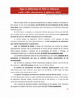
Fig. 8 Antibacterial effects of green synthesized AgNPs from H. isora
high electronegativity can easily accept electron from silver
nanoparticles. (Rodriguez-Gattorno et al. 2002).
The reducing power
Figure 7d shows the dose-dependent response for the
reducing powers of the biosynthesized AgNPs of root
extracts. Reducing power was increased consistently with
increasing the concentration of AgNPs. Surprisingly, the
AgNPs exhibited comparatively better reducing power than
standard (BHT) due to the presence of phytoconstituents in
the extracts. However, these phytoconstituents like steroidal saponins also have electron-donating antioxidant
capacity (Lin et al. 1996). This result was correlated with
biosynthesized AgNPs of Iresine herbstii (Dipankar and
Murugan 2012).
Antibacterial activity
The AgNPs exhibited good antibacterial activity against both
Gram-negative and Gram-positive bacteria (Fig. 8). But it
showed higher antibacterial activity against S. typhi and P.
123
Appl Nanosci
Fig. 9 Antibacterial activity of synthesized AgNPs against various pathogenic bacterial strains
aeruginosa (Gram negative) than B. subtilis and M. luteus
(Gram positive) (Fig. 9). This result is possible due to the
difference in the structure of the cell wall between Grampositive and Gram-negative bacteria. However, zone of
inhibition was observed less in E. coli and V. cholerae (Gram
positive), these results indicate that AgNPs show very less
antibacterial activity against these microorganisms. Peptidoglycan is composed of a thick layer of bacterial cell wall,
consisting of linear polysaccharide chains cross-linked by
short peptides thus forming more rigid structure leading to
difficult penetration of the AgNPs (Chaloupka et al. 2010).
This high bactericidal activity is certainly due to the silver
cations released from AgNPs that act as reservoirs for the
Ag? bactericidal agent (Paszek et al. 2012). Therefore,
AgNPs were widely used in antibacterial coatings in processing of medical instruments (Eby et al. 2009) and food
industries for packaging (Krishnaraj et al. 2010). The biologically synthesized AgNPs using different plant extracts
also showed a similar potent bactericidal activity
(Muthukrishnan et al. 2015; (Logeswari et al. 2013; Murugan
et al. 2014; Nithya Deva Krupa and Raghavan 2014).
Conclusion
AgNPs have emerged as a typical antimicrobial nanomaterial applied in industry, daily life, and medicine. Due to
the strong activity of AgNPs and release of Ag ions, the
biological properties and safety thereof have attracted
tremendous attentions from scientists in recent era. A
simple, stable and eco-friendly method of biosynthesizing
123
AgNPs was successfully developed using H. isora root
extract. H. isora root contains more triterpenes that play
major roles as reducing as well as capping agents for use in
synthesis of AgNPs. The extract acts as both reducing and
stabilizing agent which was confirmed by FTIR studies.
TEM and XRD reports revealed that synthesized AgNPs
were crystalline in nature with an average particle size of
30–40 nm. This biosynthesized AgNPs were found to be
multifunctional with good antioxidant activities. This
biosynthesized method facilitates best alternative for both
chemical and other physical methods. Hence, this method
can be employed in large-scale production and can be used
in many medicinal and technological applications.
Acknowledgments The work was financially supported by
University Grant Commission-Rajiv Gandhi National Fellowship
(UGC-RGNF) (No: F1-17.1/2013-14/RGNF-2013-14-SC-TAM44942. (SA-III)) University Grant Commission New Delhi, India to
the first author. We thank sophisticated analytical instrument facility
(SAIF), North-Eastern Hill University (NEHU), Shillong for accessing TEM facility. The authors wish to thank the following individuals
who provided valuable advice in the final stage of the revision process: V. Ramalingam, Research Scholar (Department of Marine science, Bharathidasan University, Tiruchirappalli, 620 024, Tamil
Nadu, India), N. Kanipandian & KS Rajkumar (Research Scholars,
Department of Animal Science, Bharathidasan University, Tiruchirappalli, 620 024, India).
Open Access This article is distributed under the terms of the
Creative Commons Attribution 4.0 International License (http://creativecommons.org/licenses/by/4.0/), which permits unrestricted use,
distribution, and reproduction in any medium, provided you give
appropriate credit to the original author(s) and the source, provide a
link to the Creative Commons license, and indicate if changes were
made.
Appl Nanosci
References
Ahmad A, Mukherjee P, Senapati S, Mandal D, Khan MI, Kumar R
(2003) Extracellular biosynthesis of silver nanoparticles using
the fungus Fusarium oxysporum. Coll Surf B Biointerfaces
28:313–318
Brand-Williams W, Cuvelier ME, Berset C (1995) Use of a free
radical method to evaluate antioxidant activity. Food Sci
Technol-LWT 28:25–30
Chaloupka K, Malam Y, Seifalian AM (2010) Nanosilver as a new
generation of nanoproduct in biomedical applications. Trends
Biotechnol 28(11):580–588
Chopra RN, Chopra IC, Handa KL, Kapoor LD (1956) Glossary of
medicinal Plants. CSIR, New Delhi, p 131
Dipankar C, Murugan S (2012) The green synthesis, characterization
and evaluation of the biological activities of silver nanoparticles
synthesized from Iresine herbstii leaf aqueous extracts. Coll Surf
B Biointerfaces 98:112–119
Eby DM, Luckarift HR, Johnson GR (2009) Hybrid antimicrobial
enzyme and silver nanoparticle coatings for medical instruments.
ACS Appl Mater Interfaces 1(7):1553–1560
Geethalakshmi R, Sarada DVL (2010) Synthesis of plant-mediated
silver nanoparticles using Trianthema decandra extract and
evaluation of their anti microbial activities. Int J Eng Sci Tech
2(5):970–975
Ghaffari-Moghaddam M, Hadi-Dabanlou R, Khajeh M, Rakhshanipour M, Shameli K (2014) Green synthesis of silver nanoparticles using plant extracts. Korean J Chem Eng 31(4):548–557
He W, Zhou YT, Wamer WG, Boudreau MD, Yin JJ (2012)
Mechanisms of the pH dependent generation of hydroxyl
radicals and oxygen induced by Ag nanoparticles. Biomaterials
33(30):7547–7555
Henglein A (1998) Colloidal silver nanoparticles: photochemical
preparation and interaction with O2, CCl4, and some metal ions.
Chem Mater 10(1):444–450
Inbathamizh L, Ponnu TM, Mary EJ (2013) In vitro evaluation of
antioxidant and anticancer potential of Morinda pubescens
synthesized silver nanoparticles. J pharm Res 6(1):32–38
Jiang H, Moon K, Zhang Z, Pothukuchi S, Wong CP (2006) Variable
frequency microwave synthesis of silver nanoparticles.
J Nanopart Res 8(1):117–124
Kalaiselvi A, Roopan SM, Madhumitha G, Ramalingam C, Elango G
(2015) Synthesis and characterization of palladium nanoparticles
using Catharanthus roseus leaf extract and its application in the
photo-catalytic degradation. Spectrochim Acta A Mol Biomol
Spectrosc 135:116–119
Kalyanasundaram GT, Doble M, Gummadi SN (2012) Production and
downstream processing of (1 ? 3)-b-D-glucan from mutant
strain of Agrobacterium sp. ATCC 31750. AMB Express 2(1):31
Kanipandian N, Kannan S, Ramesh R, Subramanian P, Thirumurugan
R (2014) Characterization, antioxidant and cytotoxicity evaluation of green synthesized silver nanoparticles using Cleistanthus
collinus extract as surface modifier. Mater Res Bull 49:494–502
Kirtikar KR, Basu BD (1993) Indian Medicinal plants, 2nd edn, vol 1,
pp 371–372. Lalit Mohan Basu, Allahabad
Krishnaraj C, Jagan EG, Rajasekar S, Selvakumar P, Kalaichelvan
PT, Mohan N (2010) Synthesis of silver nanoparticles using
Acalypha indica leaf extracts and its antibacterial activity against
water borne pathogens. Coll Surf B Biointerfaces 76(1):50–56
Kumar G, Banu GS, Murugesan AG, Rajasekara-Pandian M (2007)
Preliminary toxicity and phytochemical studies of aqueous bark
extract of Helicteres isora L. Int J Pharm 3:96–100
Kumar B, Smita K, Cumbal L, Debut A (2014) Synthesis of silver
nanoparticles using Sacha inchi (Plukenetia volubilis L.) leaf
extracts. Saudi J Boil Sci 21(6):605–609
Li P, Mao Z, Lou J, Li Y, Mou Y, Lu S, Zhou L (2011) Enhancement
of diosgenin production in Dioscorea zingiberensis cell cultures
by oligosaccharides from its endophytic fungus Fusarium
oxysporum Dzf17. Molecules 16(12):10631–10644
Lin YL, Juan IM, Chen YL, Liang YC, Lin JK (1996) Composition of
polyphenols in fresh tea leaves and associations of their oxygenradical-absorbing capacity with antiproliferative actions in
fibroblast cells. J Agric Food Chem 44:1387–1394
Logeswari P, Silambarasan S, Abraham J (2013) Ecofriendly
synthesis of silver nanoparticles from commercially available
plant powders and their antibacterial properties. Sci Iran
20(3):1049–1054
Mata R, Nakkala JR, Sadras SR (2015) Biogenic silver nanoparticles
from Abutilon indicum: their antioxidant, antibacterial and
cytotoxic effects in vitro. Coll Surf B Biointerfaces 128:276–286
Mathew PJ, Unnithan MC (1992) Search for plant having anticancer
properties used by the tribal of Wayanad, Malappuram and
Palghat districts of Kerala, India. Aryavaidyan 6(1):54–60
Molyneux P (2004) The use of the stable free radical diphenylpicrylhydrazyl (DPPH) for estimating antioxidant activity. Songklanakarin J Sci Technol 26(2):211–219
Murugan K, Senthilkumar B, Senbagam D, Al-Sohaibani S (2014)
Biosynthesis of silver nanoparticles using Acacia leucophloea
extract and their antibacterial activity. Int J Nanomed 9:2431–2438
Muthukrishnan S, Bhakya S, Kumar TS, Rao MV (2015) Biosynthesis, characterization and antibacterial effect of plant-mediated
silver nanoparticles using Ceropegia thwaitesii—An endemic
species. Ind Crops Prod 63:119–124
Nithya Deva Krupa A, Raghavan V (2014) Biosynthesis of silver
nanoparticles using Aegle marmelos (Bael) fruit extract and its
application to prevent adhesion of bacteria: a strategy to control
microfouling. Bioinorg Chem Appl 2014:1–8
Oyaizu M (1986) Studies on products of browning reactions:
antioxidative activities of products of browning reaction prepared from glucosamine. Jpn J Nutr 44:307–315
Paszek E, Czyz J, Woźnicka O, Jakubiak D, Wojnarowicz J,
Łojkowski W, Ste˛pień E (2012) Zinc oxide nanoparticles impair
the integrity of human umbilical vein endothelial cell monolayer
in vitro. J Biomed Nanotechnol 8(6):957–967
Patel K, Gadewar M, Tahilyani V, Patel DK (2012) A review on
pharmacological and analytical aspects of diosgenin: a concise
report. Nat Prod Bioprospect 2(2):46–52
Pick E, Mizel D (1981) Rapid microassays for the measurement of
superoxide and hydrogen peroxide production by macrophages
in culture using an automatic enzyme immunoassay reader.
J Immunol Methods 46(2):211–226
Ramalingam V, Rajaram R, Premkumar C, Santhanam C, Dhinesh P,
Vinothkumar S, Kaleshkumar K (2014) Biosynthesis of silver
nanoparticles from deepsea bacterium Pseudomonas aeruginosa
JQ989348 for antimicrobial, antibioflim and cytotoxic activity.
J Basic Microbiol 54:928–936
Rees DD, Palmer RM, Moncada S (1989) Role of endotheliumderived nitric oxide in the regulation of blood pressure. Proc Natl
Acad Sci USA 86:3375–3378
Rodriguez-Gattorno G, Diaz D, Rendon L, Hernandez-Segura GO
(2002) Metallic nanoparticles from spontaneous reduction of
silver (I) in DMSO. Interaction between nitric oxide and silver
nanoparticles. J Phys Chem B 106(10):2482–2487
Sands DE (1993) Introduction to crystallography. Dover, New York,
p 51
Sharma B, Purkayastha DD, Hazra S, Thajamanbi M, Bhattacharjee
CR, Ghosh NN, Rout J (2014) Biosynthesis of fluorescent gold
nanoparticles using an edible freshwater red alga, Lemanea
fluviatilis (L.) C. Ag. and antioxidant activity of biomatrix
loaded nanoparticles. Bioprocess Biosyst Eng 37(12):2559–2565
123
Appl Nanosci
Shetty P, Supraja N, Garud M, Prasad TNVKV (2014) Synthesis,
characterization and antimicrobial activity of Alstonia scholaris
bark-extract-mediated silver nanoparticles. J Nanostruct Chem
4(4):161–170
Shriram V, Kumar V, Shitole MG, Shriram V, Kumar V, Shitole MG
(2008) Indirect organogenesis and plant regeneration in
Helicteres isora L. an important medicinal plant. In Vitro Cell
Dev Biol Plant 44(3):186–193
Singh SB, Singh AK, Thakur RS (1984) Chemical constituents of
the leaves of Helicteres isora. Ind J Pharmaceu Sci
46(4):148–149
Singh S, Saikia JP, Buragohain AK (2013) A novel ‘green’ synthesis
of colloidal silver nanoparticles (SNP) using Dillenia indica fruit
extract. Coll Surf B Biointerfaces 102:83–85
Sousa A, Ferreira ICFR, Barros L, Bento A, Pereira JA (2008) Effect
of solvent and extraction temperatures on the antioxidant
potential of traditional stoned table olives ‘‘alcaparras’’. Food
Sci Technol-LWT 41:739–745
123
Suthar M, Rathore GS, Pareek A (2009) Antioxidant and antidiabetic
activity of Helicteres isora (L.) fruits. Indian J Pharm Sci
71(6):695
Venkadesh S, Reddy GD, Reddy YSR, Sathyavathy D, Reddy BM
(2004) Effect of Helicteres isora root extracts on glucose
tolerance in glucose-induced hyperglycemic rats. Fitoterapia
75:364–367
Viet Quang D, Hoai Chau N (2013) The effect of hydrothermal
treatment on silver nanoparticles stabilized by chitosan and its
possible application to produce mesoporous silver powder.
J Powder Technol 2013:1–6
Weiss J, Takhistov P, Mc Clements J (2006) Functional materials in
food nanotechnology. J Food Sci 71(9):R107–R116
Wong KKY (2012) ‘‘Silver nanoparticles in medicine: is the panacea
here’’, Nanomedicine: nanotechnology. Biol Med 8(6):935–940
Yang EJ, Kim S, Kim JS, Choi IH (2012) Inflammasome formation
and IL-1b release by human blood monocytes in response to
silver nanoparticles. Biomaterials 33(28):6858–6867
Keep reading this paper — and 50 million others — with a free Academia account
Used by leading Academics

Petra Reinke
University of Virginia

Kenneth Vecchio
University of California, San Diego

Estela Blaisten-Barojas
George Mason University

Prof. Dr. rer. nat. Mohammadamin Emami
Isfahan University of Art