Comparative Biochemistry and Physiology Part A 134 (2003) 839–846
Stressor-dependent regulation of the heat shock response in
zebrafish, Danio rerio
a,1
Susanna Airaksinena,*, Christina M.I. Rabergh
, Anna Lahtia, Annukka Kaatrasaloa,
˚
Lea Sistonenb,c, Mikko Nikinmaaa
b
a
Department of Biology, Laboratory of Animal Physiology, University of Turku, FIN-20014 Turku, Finland
˚ Akademi University, P.O. Box 123, FIN-20521 Turku, Finland
Turku Centre for Biotechnology, University of Turku, Abo
c
˚ Akademi University, Turku, Finland
Department of Biology, Abo
Received 18 November 2002; received in revised form 21 January 2003; accepted 22 January 2003
Abstract
Heat shock transcription factors (HSFs) regulate expression of heat shock proteins (Hsps). We have previously shown
that in zebrafish a unique isoform, zHSF1b, disappears concomitant with heat shock-induced Hsp70 expression. To
characterize the role of zHSF1a and zHSF1b isoforms in the regulation of the stress response in vivo, we have carried
out cadmium (10–100 mM) and copper (10–30 mM) exposures in order to specify whether the disappearance of HSF1b
is specific for heat stress. After 4-h metal exposures we analyzed the expression of hsp70, zHSF1a, zHSF1b and
metallothionein (MT) by reverse transcriptase polymerase chain reaction in zebrafish liver, gonads and gills. Although
cadmium is a known inducer of Hsps, it did not affect hsp70 expression significantly in the studied tissues. Induction
of hsp70 was observed upon copper exposure in liver and gonads, but not in gills. Neither metal affected the zHSF1ay
b ratio. Both cadmium and copper exposure caused upregulation of MT, regulator of metal homeostasis and detoxification,
confirming that the tissues were subjected to metal loads. Thus, hsp70 appears to be more weakly induced upon metal
exposure than in response to heat shock and HSF1 isoforms may participate in stressor-specific regulation of hsp70.
䊚 2003 Elsevier Science Inc. All rights reserved.
Keywords: Cadmium; Copper; Danio rerio; Heat shock; Heat shock factor; Heat shock protein; Metallothionein; Zebrafish
1. Introduction
Diverse environmental signals trigger heat shock
transcription factor (HSF)-mediated activation of
heat shock (HS) genes encoding heat shock proteins (Hsps). These stress proteins play a central
role in cell protection and repair upon stress as
*Corresponding author. Finnish Game and Fisheries
¨
Research Institute, Turku Game and Fisheries Research, Itainen
¨
Pitkakatu
3, FIN-20520 Turku, Finland. Tel.: q358-205751688; fax: q358-205-751689.
E-mail address: susanna.airaksinen@rktl.fi (S. Airaksinen).
1
Present address: Leiras Oy, Pansiontie 47, FIN-20101
Turku, Finland.
well as under certain non-stressful conditions (for
reviews, see Morimoto, 1998; Feder and Hofmann,
1999; Basu et al., 2002). Among the reported
inducers of Hsps are heavy metals (Heikkila et al.,
1982; Misra et al., 1989; Fischbach et al., 1993;
Wagner et al., 1999), which are known to induce
also another set of proteins, metallothioneins
(MTs) (Durnam and Palmiter, 1981). MTs are
involved in metal homeostasis and detoxification
(Palmiter, 1998), and they are often classified as
a specific family of stress proteins, since they are
induced not only by metals, but also by cytokines
(Karin et al., 1985; De et al., 1990), mitogens
(Imbra and Karin, 1987), and glucocorticoids
1095-6433/03/$ - see front matter 䊚 2003 Elsevier Science Inc. All rights reserved.
doi:10.1016/S1095-6433(03)00017-5
�840
S. Airaksinen et al. / Comparative Biochemistry and Physiology Part A 134 (2003) 839–846
(Karin and Herschman, 1979). However, MTs are
not structurally related to the classical members of
Hsp families. Instead, their peculiar cysteine-rich
structure is specialized for high affinity metal
binding and to serve as a protein thiol source
within a cell (for review see, Klaassen et al.,
1999).
The promoter region of HS genes contains a
conserved heat shock element (HSE), which is
responsible for transcriptional regulation via HSFbinding. It has been shown that also in fish the
HSE is occupied by HSF upon hsp70 induction
(Airaksinen et al., 1998). Among the several
members of the HSF family in vertebrates discovered to date, HSF1 is the prototype of the stressresponsive HSF (for review, see Pirkkala et al.,
2001) and the functional homologue of a single
HSF in yeast (Wiederrecht et al., 1988; Sorger
and Pelham, 1988) and fruit fly (Clos et al., 1990).
We have previously cloned HSF1 from zebrafish
and shown that two isoforms HSF1a and HSF1b
exist (Rabergh
˚
et al., 2000). Recently, an additional isoform, HSF1c, has been reported in zebrafish
(Wang et al., 2001). Interestingly, the zHSF1b
isoform disappears following HS with concomitant
induction of Hsp70, the major heat-inducible Hsp
(Rabergh
˚
et al., 2000). This observation raised the
question of the role of the zebrafish HSF1 isoforms
in the regulation of the stress response upon
exposure to stressors other than heat. Since both
Hsp and MT expression can be triggered by
common stressors, such as oxidative stress or
heavy metals (Andrews, 2000; Gosslau et al.,
2001), the present study was designed to characterize the role of zHSF1 isoforms upon metal
stress in zebrafish in vivo. Cadmium and copper
exposures were carried out in order to specify
whether the disappearance of HSF1b is stressordependent. The results suggest that the role of
HSF1 isoforms in the regulation of metal inducible
hsp70 expression is distinct from that observed
upon heat stress, where HSF1ayHSF1b ratio is
dramatically changed upon hsp70 induction
˚
(Rabergh
et al., 2000). Furthermore, high sublethal
concentrations of cadmium and copper cause only
a minor increase in the hsp70 mRNA and protein
levels in zebrafish tissues although the MT expression is readily induced.
2. Materials and methods
2.1. Animals and experimental design
Adult zebrafish (Danio rerio) were obtained
from a local aquarium store and allowed to acclimatize to laboratory conditions for a minimum of
2 weeks prior to the experiments. Fish were
maintained at 28 8C in dechlorinated carbon filtered Turku tap water (wNaqxf8.8 mgyl,
wClyxf24.8
wCd2qxf0.001
mgyl,
mgyl,
wCu2qxf0.022 mgyl, wCaCO3xf0.69 mmolyl, pH
7.0) in 12 h:12 h light–dark cycle and fed daily
(TetraMin, TetraWerke, Germany). Before the metal exposure, fish were transferred to aerated 500ml glass containers with a water temperature of 28
8C, and allowed to recover for an hour. Thereafter,
the required volume of water was substituted with
10 mM metal solution (CdCl2 and CuSO4 in
double distilled water), so that the desired concentrations were obtained. Double distilled water was
used for the controls in order to cause a similar
physical disturbance to both experimental and
control groups. According to pilot experiments the
exposure time and the concentrations showing the
maximal hsp70 induction (without recovery period) were chosen to be analyzed in detail, i.e., 4-h
exposure with 50 mM CdCl2 (5.6 mgyl) and 4-h
exposure with 20–30 mM CuSO4 (1.3–1.9 mgyl).
Each treatment was repeated 2–4 independent
times. Immediately after the 4-h exposure fish
were decapitated, and the gills, female gonads and
liver were dissected on ice.
2.2. Reverse transcriptase polymerase chain
reaction
Liver and gill samples were pooled from three
to five fish and gonad samples from one to two
fish. Tissues were homogenized on ice immediately after dissection (ULTRA-TURRAX, Ika Labortechnik, Germany) in RNAzolB solution (Tel-Test,
Inc., Friendswood, TX). Total RNA was isolated
using the RNAzolB method according to the manufacturer’s instructions. Synthesis of cDNA was
performed with 5 mg of RNA using general
oligo(dT)15-primer (Promega, Madison, WI),
together with avian myeloblastosis virus reverse
transcriptase (Finnzymes, Espoo, Finland). Subsequent amplification reactions with gene-specific
primers of zebrafish HSF1a, HSF1b, hsp70, and
hsc70 were performed as described earlier
(Rabergh
et al., 2000). The amplified products
˚
obtained were 605, 700, 457, and 412 bp, respectively. MT was amplified using primers designed
based on the zebrafish MT sequence available in
GenBank (accession no. NM_131075; forward, 59-
�S. Airaksinen et al. / Comparative Biochemistry and Physiology Part A 134 (2003) 839–846
atggatccttgcgaatg-39; reverse, 59-tcactgacaacagctgg39). The optimum annealing temperature for the
MT-primers was determined to be 47 8C with the
gradient program (HYBAID thermal cycler) and
the product size was 183 bp. In order to confirm
that the obtained sequence was MT, the product
was sequenced (ABI Prism 377, Perkin Elmer,
UK). The PCR-products were run on 1.4% TBEagarose gels, whereafter the gels were photographed under UV-light. The unsaturated bands
were quantified using Chemi-imager with
AlphaEase娃 software (Alpha Innotech Corporation, San Leandro, CA) with respect to Hsc70,
which is an evenly and ubiquitously expressed
member of hsp70 family. Generally, 3–4 independent samples were analyzed, and the statistical
significance of the differences tested with t-test.
However, due to the lack of mature female fish
among 50 mM cadmium-treated fish Hsp70 and
MT transcript levels were obtained only from one
and two individual gonad samples, respectively.
2.3. Western blot analysis
Whole cell extracts were prepared from gills,
gonads and livers by a modification of the method
described by Mosser et al. (1988). Briefly, dissected tissues, which were kept on ice throughout
the procedure, were homogenized (ULTRATURRAX) and sonicated with Branson microtip
sonicator (G. Heinemann, Ultraschalltechnik, Germany) in buffer containing 25% glycerol (vyv),
420 mM NaCl, 1.5 mM MgCl2, 0.2 mM EDTA,
20 mM HEPES, 0.5 mM phenylmethylsulfonyl
fluoride, 0.5 mM dithiotreitol. Thereafter, samples
were centrifuged for 30 min (4 8C, 14 000=g)
and the supernatants were collected and stored at
y70 8C. The protein concentration of the whole
cell extracts was determined using the Bio-Rad
protein assay according to the manufacturer’s
instructions (Bio-Rad, California, USA).
Equal amounts of protein (20 mg) were loaded
on a discontinuous SDS-polyacrylamide gel (8%),
separated and transferred to nitrocellulose
membrane (Schleicher & Schuell) using a semidry transfer apparatus (Bio-Rad). The membranes
were blocked as described earlier (Rabergh
et al.,
˚
2000). Hsp70 protein was detected using a monoclonal mouse anti-HSP70 antibody (clone 3a3,
Affinity Bioreagents, Golden, CO) diluted
1:10 000. b-Actin was detected using a mouse
anti-b-actin antibody (clone AC-15, Sigma-
841
Aldrich, Inc.) diluted 1:2000. In order to perform
enhanced chemiluminescence (Amersham Pharmacia Biotech UK Limited), the horseradish
peroxidase-conjugated rabbit anti-mouse immunoglobulin (Amersham) was used as a secondary
antibody. The signal was captured on X-ray film
and results were photographed and analyzed with
Chemi-imager (Alpha Innotech Corp.). Three separate experiments were carried out with cadmium
and two with copper.
3. Results
3.1. The ratio of zHSF1ayzHSF1b isoforms
remains unaltered following a short term cadmium
exposure
Cadmium is a potent inducer of Hsp70 as
reported in numerous studies on mammalian as
well as on fish cell cultures (Levinson et al., 1980;
Heikkila et al., 1982; Misra et al., 1989; Fischbach
et al., 1993; Steiner et al., 1998). We exposed
zebrafish to cadmium for 4 h, after which the
expression of HSF1, hsp70, and hsc70 (constitutive form of Hsp70) was analyzed by RT-PCR. In
liver and gills cadmium had no significant effect
on the hsp70 expression as determined based on
three and four independent experiments, respectively (Fig. 1a). However, when change occurred
it was upregulation of hsp70. Also in the only
gonad sample obtained at a cadmium concentration
of 50 mM increase in the hsp70 expression was
observed and the increase was evident already at
20 mM cadmium concentration (data not shown).
The absence of significant induction was further
confirmed with northern blot analysis (data not
shown).
Western blotting was used to analyze Hsp70
protein levels (Fig. 1b). In agreement with the RTPCR data, there was no detectable increase in
Hsp70 in the studied tissues when normalized
against b-actin (ns3). Again, however, in one or
two cases slight induction was observed in liver
and gonads, respectively. As a comparison, an
increased Hsp70 level in zebrafish liver was shown
after 1-h HS (9 8C above the control temperature).
As previously reported, heat-inducible isoform of
Hsp70 is detected in gills and liver, and to a lesser
extent in gonads upon HS (Rabergh
et al., 2000),
˚
indicating tissue-specificity of the temperatureinduced responses.
�842
S. Airaksinen et al. / Comparative Biochemistry and Physiology Part A 134 (2003) 839–846
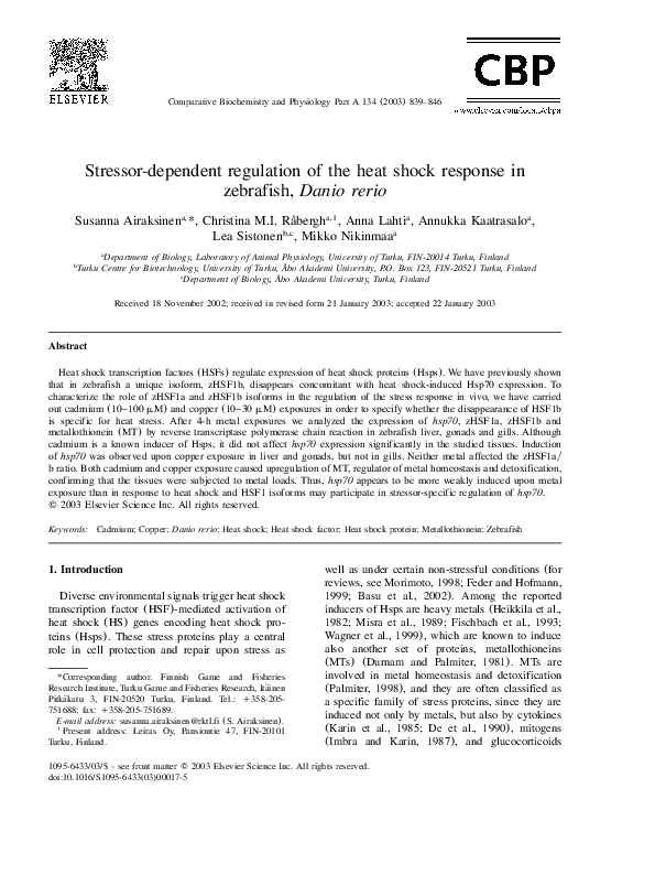
Fig. 1. (a) Expression of hsp70, two zHSF1 isoforms a and b, and hsc70 following cadmium exposure (4 h) presented by RT-PCR.
Gene-specific primers were used to study different tissues, i.e., liver, gonads and gills. The presented samples were pooled from five
fish, except 0 and 50 mM gonad samples, which were obtained from three and one female, respectively. Hsp70yHsc70 and HSF1ayb
ratios are shown below the panels. (b) Western blot analysis of zebrafish exposed to cadmium. Hsp70 (above) and b-actin (below)
were analyzed in liver, gonads and gills. The samples were pooled from four fish, except the gonad samples, which were pooled from
two fish. Heat-shocked (1 h, 37 8C) zebrafish liver sample is designated as HS. (c) Expression of MT and hsc70 presented as described
above. The presented samples were pooled from five fish, except 0 and 50 mM gonad samples, which were obtained from two and one
female, respectively. MTyHsc70 ratios are shown below the panels. (d) Dependency of MT expression on cadmium concentration in
liver (pooled from five fish) presented as described above. Cadmium concentrations are indicated above each panel and MTyHsc70
ratios are shown as in (c).
Since Hsp70 was poorly induced by cadmium,
which is generally considered as a potent inducer
of the HS response, we investigated whether the
concentrations of metal used in the exposures were
adequate to cause an upregulation of MT, a common indicator for metal exposure (Samson and
Gedamu, 1998). Cadmium, which caused only a
minor (liver and gonads) or hardly detectable
(gills) induction of hsp70, caused a prominent
induction of MT in liver (P-0.05, ns4) and gills
(P-0.05, ns4) at 50 mM concentration (Fig. 1c).
As shown in Fig. 1d, MT levels increased in a
dose-dependent manner. In contrast, the expression
level of MT in gonads was unaffected (ns2) (Fig.
1c).
3.2. Hsp70 is more efficiently induced than MT by
copper
We analyzed the expression of HSF1, hsp70,
and hsc70 following a 4-h exposure to copper, an
essential metal, which is a cofactor of many
enzymes and is also known to induce MTs (Durnam and Palmiter, 1981; Nieminen and Lemasters,
1996). The hsp70 expression was moderately
increased at copper concentrations of 25 mM in
liver (P-0.1, ns4) and gonads (P-0.06, ns3),
whereas the expression in gills remained unaffected (ns3)(Fig. 2a). Similar to the cadmium treatments, no change was observed in the ratio of
HSF1 isoforms in any of the tissues (Fig. 2a).
Western blot analysis suggests comparable Hsp70
increase in liver and gonads following copper
exposure (ns2) (Fig. 2b). As a comparison, a
markedly elevated Hsp70 protein level in liver
induced by HS is shown in Fig. 2b. To avoid
further stress for experimental fish due to handling
no recovery period after the exposure was allowed.
This may limit the degree of induction at the
protein level.
Next we studied whether the concentrations of
copper used in this study were sufficient to induce
the expression of MT. In the liver, a slight increase
in MT expression (P-0.1, ns4) was observed at
�S. Airaksinen et al. / Comparative Biochemistry and Physiology Part A 134 (2003) 839–846
843
study upon heat-inducible hsp70 induction
(Rabergh
et al., 2000). Accordingly, the HSF1b
˚
isoform disappeared gradually at 35–37 8C (upper
panel in Fig. 3). The pattern of expression upon
metal exposure was, however, distinct from that
observed upon heat stress (lower panel in Fig. 3).
Neither of the metals affected the HSF1ayb ratio
even though the target gene was upregulated by
copper (Fig. 2). In gonads, zHSF1a and zHSF1b
were equally expressed regardless of the treatment,
and in the liver both isoforms were present at
control temperature and both in cadmium and
copper-treated animals, whereas zHSF1b disappeared during HS. In gills zHSF1a was a dominant
isoform under all conditions (Fig. 1a and Fig. 2a).
4. Discussion
4.1. Cadmium and copper as stress inducers
Fig. 2. (a) Expression of hsp70, two zHSF1 isoforms a and b,
and hsc70 following copper exposure (4 h) presented by RTPCR. Gene-specific primers were used to study different tissues, i.e., liver, gonads and gills. The samples were pooled
from five, two and five fish, respectively. Hsp70yHsc70 and
HSF1ayb ratios are shown below the panels. (b) Western blot
analysis of zebrafish exposed to copper. Hsp70 (above) and bactin (below) were analyzed in liver, gonads and gills. The
samples were pooled from four fish, except the gonad samples,
which were pooled from one, two and four fish, respectively.
Heat shocked (1 h, 37 8C) zebrafish liver sample is designated
as HS. (c) Expression of MT and hsc70 presented as described
above. The liver sample was pooled from five, the gonad sample from two, and the gill sample from five fish. Copper concentrations are indicated above each panel and MTyHsc70
ratios are shown as in Fig. 1c.
concentration of 25 mM suggesting an adequate
copper load (Fig. 2c). Indeed, higher concentrations of copper (035 mM) proved to be lethal to
the fish, indicating the severity of the stress (data
not shown). The tissue-specific response was
reflected in the observation that MT induction was
reversed in gonads (P-0.05, ns4) and absent in
gills (ns3) (Fig. 2c).
3.3. Stressor-specific regulation of the heat shock
response by zHSF1 isoforms
A dramatic change in the ratio of HSF1 isoforms
was observed in zebrafish liver in our previous
Surprisingly, well known inducers of HS
response proved to be only modest inducers of
Hsp70 in adult zebrafish liver, gonad and gill
tissue. The observed induction of hsp70 following
HS could be mediated by stimulating the activatory
effect of zHSF1a andyor eliminating the inhibitory
effect of zHSF1b. Neither cadmium nor copper
caused a change in the zHSF1ayb ratio. If a change
in the HSF isoform ratio is indeed required for
full hsp70 induction, the unaltered ratio could be
reflected in the diminished Hsp70 induction in
response to metal treatments, when compared to
Fig. 3. Comparison of zHSF1a and zHSF1b expression after 1
h HS (upper panel) or after 4 h cadmium and copper exposure
(lower panels). HS temperatures (33, 35, or 37 8C) and concentrations of cadmium and copper treatments (28 8C) are indicated above the panels. The ratios of HSF1ayHSF1b expression
following treatments as obtained by image analysis are shown
below the panels. The presented samples were pooled from five
fish.
�844
S. Airaksinen et al. / Comparative Biochemistry and Physiology Part A 134 (2003) 839–846
HS. The binding of zHSF1 to the intact hsp70
promoter was not analyzed in current study. Wang
et al. have, however, reported enhanced binding of
in vitro translated zHSF1a and zHSF1c to HSE at
elevated temperature (Wang et al., 2001).
An explanation for the profound induction of
MT concurrent with the minor HS response could
be that the metal load was sequestered by MTs
(Foulkes and McMullen, 1986). However, it has
been shown that at least in zebrafish gills the
cadmium-binding capacity, including MT-binding,
is exceeded at as low as 15 nM concentration of
cadmium, whereupon influx into circulation is
drastically increased (Wicklund, 1996). Therefore,
current results may also reflect variability of different tissues in adjusting the response to a given
stressor. The metal inducible response appeared to
follow tissue-specific patterns, as observed before
with heat stress (Rabergh
et al., 2000). It is
˚
noteworthy that the sublethal metal concentrations
used in this study correspond to the concentrations
used in cell cultures, where they have been shown
to induce the HS response (Heikkila et al., 1982;
Misra et al., 1989; Ryan and Hightower, 1994;
Croute et al., 2000). Furthermore, the concentrations used were close to the upper limits tolerated
by zebrafish in vivo. A recent study by Blechinger
et al. (2002) has monitored the cadmium-induced
hsp70 expression in early larvae (80 h) of zebrafish. Interestingly, both endogenous hsp70 expression and reporter gene expression under hsp70
promoter showed dose-dependent increase in gills
(00.2 mM) and liver (0125 mM), which adds
the life-stage specificity to the list of variability
creating factors.
MT protects the animal most effectively against
cadmium toxicity, when compared to other metals
such as copper, zinc, iron, lead, mercury, or arsenite (Park et al., 2001). Also, cadmium has been
shown to be a better inducer of MT in bovine
chondrocytes and mouse tissues compared to copper (Durnam and Palmiter, 1981; Zafarullah et al.,
1993). Furthermore, when the metal regulatory
element (MRE) driven luciferase activity was
measured in zebrafish cell line, ZEM2S, upon a
number of metal treatments, the inducibility was
lowest with copper and highest with cadmium
(Carvan et al., 2000). Observations above correspond to the difference in MT induction observed
in this study between cadmium and copper treatment. The tissue distribution of metals was not
measured in the present study. However, depending
on the cadmium-sensitivity of the fish species liver,
kidney and gills are known to accumulate most of
the cadmium in the course of time (Norey et al.,
2002).
4.2. Is there a signaling network connecting heat
and metal stress?
A common regulatory element in the promoter
of HS genes, HSE, is also found in the superoxide
dismutase (SOD1) promoter, where it is occupied
upon treatment by two distinct stressors, paraquat,
a Oy
2 generating agent, and HS (Yoo et al., 1999).
The communication in this case appears to occur
at the level of transcription, and may be mediated
through the same signaling molecule, i.e., superoxide. This suggests an involvement of redox
reactions in the process. Interestingly, many metals,
such as copper, are capable of changes in valency
thus affecting the cellular redox status. Both Hsp70
and MT promoters have been studied intensively
in order to find out whether the regulatory regions
of these genes might also have common elements.
It appears, however, that this is not the case.
Although the MT gene expression is also regulated
at the transcriptional level, the MRE recruited by
a specific metal transcription factor, MTF-1, upon
activation is distinct from HSE (Stuart et al., 1985;
Radtke et al., 1993; Olsson et al., 1995; Dalton et
al., 1997; Samson et al., 2001; Chen et al., 2002).
4.3. Stress response at cellular vs. organismic level
Numerous studies performed on cell cultures as
well as general statements about the HS response
suggest that the Hsps are prominently induced by
heavy metals. Based on our results with cadmium
where MTs were induced in vivo in the virtual
absence of HS response, this statement may be too
simplistic in biologically relevant context. The
nature of the stressor and its signaling pathway
within each tissue of an organism should therefore
be taken into account when estimating the biological significance of a specific type of stress to its
target.
In conclusion, in the studied tissues of adult
zebrafish Hsp70 appears to be weakly, if at all
induced upon metal exposures, compared to the
marked induction observed in response to heat
stress. Since the zHSF1-isoform ratio is markedly
changed as a response to elevated temperatures,
but remains unaffected by metals, this stressor-
�S. Airaksinen et al. / Comparative Biochemistry and Physiology Part A 134 (2003) 839–846
specific response of HSF may be an important
determinant in the induction of HS response in
intact animal tissues when exposed to different
stressors. This study highlights the great complexity of the stress response, which becomes apparent
only when experiments are conducted at the organismic level instead of isolated cells in culture.
Acknowledgments
This work was supported by the Academy of
Finland, projects 40830, 42186 and 50748.
References
Airaksinen, S., Rabergh,
C.M.I., Sistonen, L., Nikinmaa, M.,
˚
1998. Effects of heat shock and hypoxia on protein synthesis
in rainbow trout (Oncorhynchus mykiss) cells. J. Exp. Biol.
201, 2543–2551.
Andrews, G.K., 2000. Regulation of metallothionein gene
expression by oxidative stress and metal ions. Biochem.
Pharmacol. 59, 95–104.
Basu, N., Todgham, A.E., Ackerman, P.A., et al., 2002. Heat
shock protein genes and their functional significance in fish.
Gene 295, 173–183.
Blechinger, S.R., Warren Jr., J.T., Kuwada, J.Y., Krone, P.H.,
2002. Developmental toxicology of cadmium in living
embryos of a stable transgenic zebrafish line. Environ.
Health Perspect. 110, 1041–1046.
Carvan III, M.J., Solis, W.A., Gedamu, L., Nebert, D.W., 2000.
Activation of transcription factors in zebrafish cell cultures
by environmental pollutants. Arch. Biochem. Biophys. 376,
320–327.
Chen, W.Y., John, J.A., Lin, C.H., Chang, C.Y., 2002. Molecular cloning and developmental expression of zinc finger
transcription factor MTF-1 gene in zebrafish, Danio rerio.
Biochem. Biophys. Res. Commun. 291, 798–805.
Clos, J., Westwood, J.T., Becker, P.B., Wilson, S., Lambert,
K., Wu, C., 1990. Molecular cloning and expression of a
hexameric Drosophila heat shock factor subject to negative
regulation. Cell 63, 1085–1098.
Croute, F., Beau, B., Arrabit, C., et al., 2000. Pattern of stress
protein expression in human lung cell-line A549 after shortor long-term exposure to cadmium. Environ. Health Perspect. 108, 55–60.
Dalton, T., Paria, B.C., Fernando, L.P., Huet-Hudson, Y.M.,
Dey, S.K., Andrews, G.K., 1997. Activation of the chicken
metallothionein promoter by metals and oxidative stress in
cultured cells and transgenic mice. Comp. Biochem. Physiol.
Part B 116, 75–86.
De, S.K., McMaster, M.T., Andrews, G.K., 1990. Endotoxin
induction of murine metallothionein gene expression. J.
Biol. Chem. 265, 15267–15274.
Durnam, D.M., Palmiter, R.D., 1981. Transcriptional regulation
of the mouse metallothionein-I gene by heavy metals. J.
Biol. Chem. 256, 5712–5716.
Feder, M.E., Hofmann, G.E., 1999. Heat-shock proteins,
molecular chaperones, and the stress response: evolutionary
and ecological physiology. Annu. Rev. Physiol. 61, 243–282.
845
Fischbach, M., Sabbioni, E., Bromley, P., 1993. Induction of
the human growth hormone gene placed under human hsp70
promoter control in mouse cells: a quantitative indicator of
metal toxicity. Cell Biol. Toxicol. 9, 177–188.
Foulkes, E.C., McMullen, D.M., 1986. Endogenous metallothionein as determinant of intestinal cadmium absorption: a
reevaluation. Toxicology 38, 285–291.
Gosslau, A., Ruoff, P., Mohsenzadeh, S., Hobohm, U., Rensing, L., 2001. Heat shock and oxidative stress-induced
exposure of hydrophobic protein domains as common signal
in the induction of hsp68. J. Biol. Chem. 276, 1814–1821.
Heikkila, J.J., Schultz, G.A., Iatrou, K., Gedamu, L., 1982.
Expression of a set of fish genes following heat or metal
ion exposure. J. Biol. Chem. 257, 12000–12005.
Imbra, R.J., Karin, M., 1987. Metallothionein gene expression
is regulated by serum factors and activators of protein kinase
C. Mol. Cell Biol. 7, 1358–1363.
Karin, M., Herschman, H.R., 1979. Dexamethasone stimulation
of metallothionein synthesis in HeLa cell cultures. Science
204, 176–177.
Karin, M., Imbra, R.J., Heguy, A., Wong, G., 1985. Interleukin
1 regulates human metallothionein gene expression. Mol.
Cell Biol. 5, 2866–2869.
Klaassen, C.D., Liu, J., Choudhuri, S., 1999. Metallothionein:
an intracellular protein to protect against cadmium toxicity.
Annu. Rev. Pharmacol. Toxicol. 39, 267–294.
Levinson, W., Oppermann, H., Jackson, J., 1980. Transition
series metals and sulfhydryl reagents induce the synthesis
of four proteins in eukaryotic cells. Biochim. Biophys. Acta
606, 170–180.
Misra, S., Zafarullah, M., Price-Haughey, J., Gedamu, L.,
1989. Analysis of stress-induced gene expression in fish
cell lines exposed to heavy metals and heat shock. Biochim.
Biophys. Acta 1007, 325–333.
Morimoto, R.I., 1998. Regulation of the heat shock transcriptional response: cross talk between a family of heat shock
factors, molecular chaperones, and negative regulators.
Genes Dev. 12, 3788–3796.
Mosser, D.D., Theodorakis, N.G., Morimoto, R.I., 1988. Coordinate changes in heat shock element-binding activity and
HSP70 gene transcription rates in human cells. Mol. Cell
Biol. 8, 4736–4744.
Nieminen, A.-L., Lemasters, J.J., 1996. Hepatic injury by
metal accumulation. In: Chang, L.W. (Ed.), Toxicology of
Metals. CRC Press Inc, pp. 887–901.
Norey, C.G., Cryer, A., Kay, J., 2002. A comparison of
cadmium-induced metallothionein gene expression and
Me2q distribution in the tissues of cadmium-sensitive
(Rainbow trout; Salmo gairdneri) and tolerant (Stone loach;
Noemacheilus barbatulus) species of freshwater fish. Comp.
Biochem. Physiol. Part C 97, 221–225.
Olsson, P.E., Kling, P., Erkell, L.J., Kille, P., 1995. Structural
and functional analysis of the rainbow trout (Oncorhyncus
mykiss) metallothionein-A gene. Eur. J. Biochem. 230,
344–349.
Palmiter, R.D., 1998. The elusive function of metallothioneins.
Proc. Natl. Acad. Sci. 95, 8428–8430.
Park, J.D., Liu, Y., Klaassen, C.D., 2001. Protective effect of
metallothionein against the toxicity of cadmium and other
metals (1). Toxicology 163, 93–100.
�846
S. Airaksinen et al. / Comparative Biochemistry and Physiology Part A 134 (2003) 839–846
Pirkkala, L., Nykanen, P., Sistonen, L., 2001. Roles of the heat
shock transcription factors in regulation of the heat shock
response and beyond. FASEB J. 15, 1118–1131.
Radtke, F., Heuchel, R., Georgiev, O., et al., 1993. Cloned
transcription factor MTF-1 activates the mouse metallothionein I promoter. EMBO J. 12, 1355–1362.
Ryan, J.A., Hightower, L.E., 1994. Evaluation of heavy metal
ion toxicity in fish cells using a combined stress protein
and cytotoxicity assay. Environ. Toxicol. Chem. 13,
1231–1240.
Rabergh,
C.M.I., Airaksinen, S., Soitamo, A., et al., 2000.
˚
Tissue-specific expression of zebrafish (Danio rerio) heat
shock factor 1 mRNAs in response to heat stress. J. Exp.
Biol. 203, 1817–1824.
Samson, S.L., Gedamu, L., 1998. Molecular analyses of
metallothionein gene regulation. Prog. Nucleic Acid Res.
Mol. Biol. 59, 257–288.
Samson, S.L., Paramchuk, W.J., Gedamu, L., 2001. The rainbow trout metallothionein-B gene promoter: contributions
of distal promoter elements to metal and oxidant regulation.
Biochim. Biophys. Acta 1517, 202–211.
Sorger, P.K., Pelham, H.R.B., 1988. Yeast heat shock factor is
an essential DNA-binding protein that exhibits temperaturedependent phosphorylation. Cell 54, 855–864.
Steiner, E., Kleinhappl, B., Gutschi, A., Marth, E., 1998.
Analysis of hsp70 mRNA levels in HepG2 cells exposed to
various metals differing in toxicity. Toxicol. Lett. 96–97,
169–176.
Stuart, G.W., Searle, P.F., Palmiter, R.D., 1985. Identification
of multiple metal regulatory elements in mouse metallothionein-I promoter by assaying synthetic sequences. Nature
317, 828–831.
Wagner, M., Hermanns, I., Bittinger, F., Kirkpatrick, C.J.,
1999. Induction of stress proteins in human endothelial cells
by heavy metal ions and heat shock. Am. J. Physiol. 277,
L1026–L1033.
Wang, G., Huang, H., Dai, R., Lee, K.Y., Lin, S., Mivechi,
N.F., 2001. Suppression of heat shock transcription factor
HSF1 in zebrafish causes heat-induced apoptosis. Genesis
30, 195–197.
Wicklund, G.A., 1996. The concentration dependency of branchial intracellular cadmium distribution and influx in the
zebrafish (Brachydanio rerio). Aquat. Toxicol. 35, 47–48.
Wiederrecht, G., Seto, D., Parker, C.S., 1988. Isolation of the
gene encoding the S. cerevisiae heat shock transcription
factor. Cell 54, 841–853.
Yoo, H.Y., Chang, M.S., Rho, H.M., 1999. The activation of
the rat copperyzinc superoxide dismutase gene by hydrogen
peroxide through the hydrogen peroxide-responsive element
and by paraquat and heat shock through the same heat
shock element. J. Biol. Chem. 274, 23887–23892.
Zafarullah, M., Su, S., Gedamu, L., 1993. Basal and inducible
expression of metallothionein and heat shock protein 70
genes in bovine articular chondrocytes. Exp. Cell Res. 208,
371–377.
�
 Mikko Nikinmaa