http://dx.doi.org/10.5935/0100-4042.20160114
Quim. Nova, Vol. 39, No. 8, 914-918, 2016
Artigo
INTERACTION OF CHALCONES WITH CT-DNA BY SPECTROPHOTOMETRIC ANALYSIS AND
THEORETICALSIMULATIONS
Ximena Zarateb, Eduardo Schottc, Carlos A. Escobard, Roberto Lopez-Castroe, Cesar Echeverriaa,g, Leonor Alvarado-Sotoa,f
and Rodrigo Ramirez-Taglea,*
a
Universidad Bernardo O’Higgins, Facultad de Ingenieria, Avenida Viel 1497, Santiago, Chile.
b
Instituto de Ciencias Químicas Aplicadas, Facultad de Ingeniería, Universidad Autónoma de Chile, Av. Pedro de Valdivia 641,
Santiago, Chile
c
Departamento de Química Inorgánica, Facultad de Química, Pontificia Universidad Católica de Chile. Avda. Vicuña Mackenna
4860, Santiago, Chile
d
Universidad Andres Bello, Departamento de Ciencias Químicas, Republica 275, Santiago, Chile
e
Universidad Bernardo O´Higgins, Facultad de Salud, Deporte y Recreación, Escuela de Kinesiología, General Gana 1780,
Santiago, Chile
f
Universidad Finis Terrae, Santiago, Chile
g
Universidad Bernardo O’Higgins, Laboratorio de Bionanotecnologia, General Gana 1780, Santiago, Chile
Recebido em 11/12/2015; aceito em 27/04/2016; publicado na web em 08/07/2016
Chalcones are open chain molecules precursors of flavonoids and isoflavonoids, found spread in edible plants. Because they are
easily accessible trough Claisen Shmidt condensation, a great variety of derivatives are available. They have also shown potential
in pharmacological and biological applications. It is known that chalcone derivatives display a role in the treatment of complex
diseases such as cancer, among others, where the DNA is considered as the target for the action of these kinds of compounds.
This action is commonly explained as the inhibition of the DNA replications and transcriptions through interactions. However, not
conclusive associations between these DNA-Drug interactions and toxicity have been found. This research focuses on the capacity of
a chalcone`s family to interact with DNA. Therefore, the binding constants for each compounds with Calf Thymus DNA [CT-DNA]
were determined by spectrophotometric titration at room temperature. In addition, the effect of increasing the chalcone`s concentration
over the relative viscosity of CT-DNA at room temperature was assessed. On the other hand, with the aim to find the optimal DNAchalcone configurations, as well as consistently predict their binding, a computational work was undertaken. To accomplish these goals
within a reasonable time framework, an empirical scoring function (AScore) and a docking engine (ShapeDock) were performed using
the ArgusLab package. The results of viscosity and docking measurement provided structural insights which suggest that chalcones
bind with DNA via interaction as well as intercalation. The presence of interactions is also evidenced by the spectrophotometric study
which showed luminescence quenching of the chalcones upon interaction with CT-DNA.
Keywords: chalcones; DNA; docking; antitumoral; spectrophotometric analysis.
INTRODUCTION
Chalcones are open chain molecular systems precursors of flavonoids1 and isoflavonoids, present frequently in edible plants.
Structural modification of chalcones has allowed to obtain derivatives which display potential and worthy applications in pharmacological and biological areas like new medicinal agents with improved
properties, such as higher potency and lesser toxicity.2
This wide range of biological activities associated with many
chalcone derivatives, has stimulated interest in the development of
synthetic strategies aimed to the synthesis of heterocyclic systems
starting from chalcones. Besides of the implementation of efficient
methodologies for their obtaining, researches have focused also on
the study of their reactivity and the assessment of their possible
biological activities.3–5
Particularly in the pharmaceutical field, chalcone derivatives have
found application on the treatment of different important diseases
such as cancer, among others.6–10
DNA is thought to be the main target of antitumoral drugs, and
the binding between cisplatin complexes and DNA targets have been
extensively studied.11,12 Authors have shown that DNA interactions
*e-mail: rramirez@ubo.cl
with several compounds affect the replication process and hence this
inhibits the growth of the tumor cells, this means antitumor effects.
This effect has been the basis for designing new and more efficient
antitumor drugs. Moreover, their effectiveness depends on the mode
and affinity of their binding ability to the DNA strands.13
The interactions between DNA and drugs has been considered as
the main aspect that govern the DNA replications and transcription in
cancer cells.14 It should be considered though, that not deep studies of
DNA-Drug interactions and their toxicity have been performed.15–17
This research focuses on the capacity of a chalcone’s family
(Figure 1) to interact with DNA. Therefore, the binding constants
for the chalcone complexes with CT-DNA were determined by
spectrophotometric titration at room temperature. These procedures
were performed through the stepwise addition of a CT-DNA solution to a chalcone solution. In addition, the effect of the increasing
chalcone’s concentration over the relative viscosity of CT-DNA at
room temperature was assessed.18,19
EXPERIMENTAL
Docking
The docking procedure is envisaged as a complex optimization
�Interaction of Chalcones with CT-DNA by spectrophotometric analysis and theoreticalsimulations
Vol. 39, No. 8
915
2,5,4´-trimethoxy-2’-hydroxy-chalcone (4)9 obtained as orange crystals (75%) mp. 107 - 108 °C and 4’-methoxy-2,4-dichloro-chalcone
(5)26 obtained as white crystals (69%), mp. 136.1 - 137.4 °C.
Cell culture
C1
C2
C3
C4
C5
2´
OH
OH
H
OH
H
4´
OCH3
H
H
OCH3
OCH3
5´
H
H
Br
H
H
2
3
4
5
OCH3 OCH3
H
H
OCH3
H
OCH3
H
H
OCH3 OCH3
H
OCH3
H
H
OCH3
H
H
Cl
H
6
H
H
H
H
Cl
Figure 1. Chemical structures of the studied chalcones
or an exhaustive search process since it involves many degrees
of freedom. The ultimate goal is to find the optimal ligand/DNA
configurations, and to predict their binding free energy. To computationally accomplish this key objective within a reasonable time
framework, an empirical scoring function (AScore) and a docking
engine (ShapeDock) were employed in the ArgusLab program.20
The AScore is based on the deconvolution of the total DNA-ligand
binding free energy into different components:
∆Gbinding = ∆GvdW + ∆Ghydrophobic + ∆GH-bond + ∆GH-bond(chg) +
∆Gdeformation + ∆G°
The dissected terms account for the van der Waals interaction
between the ligand and the DNA (∆GvdW), the hydrophobic effect
(∆Ghydrophobic), the hydrogen bonding between the ligand and the protein
(∆Ghydrophobic), the hydrogen bonding involving charged donor and/or
acceptor groups (∆GH-bond(chg)), the deformation effect (∆Gdeformation),
and the effects of the translational and rotational entropy loss in the
bidding process (∆G°).
The 3D structures of chalcones were first constructed using
Arguslab , then were optimized using Austin Model 1 [AM1] , 200
maximum iteration , followed by conjugate gradient minimization to
a RMS energy gradient of 0,01 kcal/mol.21 Candidate conformations
of Chalcones interacting to their target structures, the DNA complex,
were proposed using Arguslab.22,23 Here, docking was carried out with
a set of Chalcones. (Figure 1)
Chemicals
Synthesis of Chalcones, general methodology: Chalcones were
prepared by adding dropwise a solution of the corresponding substituted benzaldehyde, (7.34 mmol in ethanol, 20 mL) to a stirred mixture
of 2-hydroxyacetophenone solution (7.34 mmol, in ethanol, 20 mL)
and potassium hydroxide solution (2 g in 10 mL distilled water).
The mixture was allowed to react overnight, and then, distilled water
(200 mL) were added and neutralized with hydrochloric acid. Then,
the mixture was extracted four times with ethyl acetate (50 mL). The
combined organic fractions were concentrated in vacuum, dissolved
in ethanol, and allowed to crystallize.
We have previously described the synthesis of the following
compounds: 2,3,4’-trimethoxy-2’-hydroxy-chalcone (1)1 which
was obtained as orange crystals (58%), mp. 129–130°C; 2,4-dimethoxy-2’-hydroxy-chalcone (2)1,7,24 obtained as yellow crystals
(58%), mp. 107 – 109.8 °C; 3´-bromo-3,4-dimethoxy-chalcone (3)25
obtained as yellow crystals (73%), mp.117–120 °C; The following
compounds were prepared as previously reported in the literature.
Spectroscopic data were identical to those previously reported:
HUVEC-derived endothelial cell line, (EA.hy926) was kindly
provided by C-J Edgell and was grown in Dulbecco´s modified Eagle
Medium (DMEM)-low glucose (GIBCO) supplemented with 10%
heat-inactiva fetal bovine serum (FBS), 2 mmol L-1 and 50 U mL-1
penicilin/streptomycin (Sigma).
Human hepatocellular carcinoma HepG2 cells (American Type
Culture Collection HB-8065) were grown in monolayer culture
in DMEM-high glucose with 10% FBS and antibiotic-antimicotic
(GIBCO). All cell cultures were grown at 37 °C in a 5%: 95% CO2:
air atmosphere controlled.
Cell viability assay
Cell viability was evaluated using the 3-(4,5-dimethylthiazol-2-yl)-2,5-diphenyltetrazolium bromide (MTT) colorimetric assay
(Invitrogen, Eugene, Oregon, USA). Cell viability was quantified by
the amount of MTT reduction27 HepG2 and EA.hy926 were exposed
to different concentrations of chalcones for 48 hours. After treatment
cells were co-incubated with MTT (0.5 mg mL) for 4 hours, and then
solubilized with an acidified (0.04 N HCl) isopropanol/dimethyl sulfoxide (DMSO) solution. Optical density was measured at 540 nm.
All experiments were performed as triplicates. Data were expressed
as percentage of survival cell. Cell survival (%) data were plotted
and adjusted to a sigmoidal best-fit curve, where: IC50 is the chalcone
concentration to reach the half-maximal cell survival.
Interaction of chalcones with CT-DNA by spectrophotometric
titration
Absorption spectroscopy is a useful tool to study the binding of
drugs to DNA. Increments in DNA concentration result in shifts of
absorption bands, which denote an effect of hyperchromism, resulting
from a direct interaction between CT-DNA and the chalcones. These
changes are similar to those small molecules that bind to double-stranded DNA through noncovalent interactions28,29
In this sense, the binding constants for the chalcone compounds
with CT-DNA were determined by absorption titration at room
temperature through the stepwise addition of a CT-DNA solution
(10 µL, ∼4×10⁻⁴ mol L-1) to a solution of each chalcone (2 mL
of (1) 4.8×10⁻⁵ mol L-1, (2) 4.8×10⁻⁵ mol L-1, (3) 5.6×10⁻⁵ mol L-1,
(4) 4.8×10⁻⁵ mol L-1 and (5) 4.8×10⁻⁵ mol L-1) in buffer Tris/HCl and
NaOH adjusted to pH 7.39.
UV-vis absorption spectra were recorded between 190-1000 nm
and the titration was finished when the intensity of those two bands
did not change significantly upon further addition of CT-DNA. The
binding constant Kb was calculated by using the Scatchard equation:
(1)
where [DNA] is the concentration of DNA, εa is the molar absorption
coefficient of the complex chalcone-DNA at given DNA concentration, εf is the molar absorption coefficient of the solution of the free
chalcone and εb is the molar absorption coefficient of the chalcone
when fully bound to DNA. A plot of [DNA]/[εa-εf] vs [DNA] gives
1/[εb-εf] as the angular coefficient. The Kb is determined by a ratio
between the angular and the linear coefficients.
�916
Zarate et al.
Viscosity
Hydrodynamic methods have been employed to test the binding
mode of DNA to agents. In this sense, viscosity changes provide
experimental advantages since viscosity is sensitive to length changes of DNA and the measures can reliably distinguish intercalation
from groove binding. When intercalation is present, a planar ligand
fragment is placed between adjacent base pairs, which induces lengthening of the helix. These interactions that result in the increase of
the DNA length generate an increase of viscosity. On the other hand,
a groove binder, typically carries out subtle changes and the DNA
remains in the unperturbed form and not increase of the DNA length
is observed, therefore this binding mode shows no increase of the
viscosity of the DNA solutions.18,19
In the herein work, the effect of the increasing concentration of
chalcones over the relative viscosity of CT-DNA at room temperature
was measured employing a viscosimeter Anton Par Lovis 2000M.
The values for relative specific viscosity (η/η0)1/3, where η0 is the
specific viscosity contributions of DNA in absence of compound
and η is the viscosity in the presence of the complex, were plotted
against [complex]/[DNA] where [complex] is the concentration of
the chalcones and [DNA] is the concentration of CT-DNA.
RESULTS AND DISCUSSION
The fact of inhibiting the replication process of DNA by action of
drugs is the motivation of designing new and more efficient antitumor
drugs. Moreover, their effectiveness depends on the mode and affinity
of their binding ability to the DNA strands.13 It is for this reason that
we employed two cell lines, which have different DNA replication
rate, where HepG2 cells have a high rate of replication with respect
to EA.hy296 cells. The interactions of chalcones with DNA may have
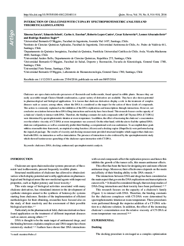
Figure 2. Molecular simulations of chalcones with DNA
Quim. Nova
Table 1. Relationship between IC50 [µM] ; HepG2 , EAhy926 , Kb and ∆G
[kcal/mol]
EAhy.926
HepG2
Chalcone
IC50
[µmol L-1]
IC50
[µmol L-1]
1
7.66
2
dG
[kcal mol-1]
Kb
10.64
-4.06
2.33E+03
10.73
31.26
-4.06
2.33E+04
3
3.91
8.27
-4.04
3.33E+04
4
12.52
21.40
-3.98
3.75E+03
5
11.92
26.18
-4.13
2.58E+04
a crucial relevance in its induction of tumor cell death. Our results
showed a higher cytotoxic effect of chalcones in EA.hy296 cells
compared to HepG2 cells (Table 1).
This selectivity of chalcones could be explained by a lower rate of
DNA replication observed in EA.hy296 cells, however both lines are
tumor and have low IC50, experiments in primary cultures of normal
cells are necessary to determine their selectivity only to tumor cells.
The IC50 and Kd values for the chalcone’s family studied here were
compared in both cell lines. To compare the effect of chalcones on
HepG2 and EA.hy296 cells, a correlation between IC50 and Kd
was established (Table 1). The docking computations suggest that
the chalcones does interact with DNA via intercalation and that the
chalcones exhibits affinity for double stranded DNA as shown in
Figure 2, such interaction with the nucleic acids could inhibit cellular
DNA synthesis during DNA replication.
According to this docking experiment, the complexes reasonably
bind with DNA. The minimum energy obtained for a docked structure
(Figure 2) suggest that the best possible conformation of the ligand
�Vol. 39, No. 8
Interaction of Chalcones with CT-DNA by spectrophotometric analysis and theoreticalsimulations
interaction, is mainly through the aromatic ring being inside the
DNA strand. It has been observed that the complex is stabilized by
electrostatic hydrogen bonding with DNA bases, in addition to van
der Waal’s and stacking–bond interactions between electron deficient
chalcone ring and purine–pyrimidine bases. The binding energy
values are presented in Table 1.
Chalcones can bind with double-stranded DNA in various binding
modes on the basis of its structure. However, hypochromic effect
could be attributed to the stacking interaction between the aromatic
rings of the ligand framework and DNA base pairs as well. The
hypochromism and bathochromic shifts may commonly vary in consistence with the strength of intercalative interaction of the complex
with DNA helix as well as overall conformation of the DNA. The
917
intrinsic binding constant of the chalcones with DNA was measured
as Kb (see Methods). The low value of the binding constant obtained
here (Kb = C1 2.33×10³, C2 2.33×10⁴, C3 3.33×10⁴, C4 3.75×10³,
C5 2.58×10⁴) suggests that the chalcones interacts with DNA double
strand in an intercalated manner (Figure 3).
On the other hand, taking into account the viscosity measurements, on increasing the amounts of C4>C5>C1>C2>C3 bound to
DNA, the relative viscosity of the DNA increases steadily (Figure 4).
The observed linear increase in viscosity as a function of chalcone
content for all compounds across the range of the relation of chalcone-DNA concentrations, suggests the fact that the interaction of the
compounds with the DNA leads to increase the length of the DNA
chains, which is indicative of the classical intercalation model.18,19
Figure 3. Electronic titration spectra of chalcones with CD-DNA , a) C1, b) C2 , c) C3 , d) C4 , e) C5
�918
Zarate et al.
Finally, the results of UV-vis spectroscopy, viscosity and docking
measurement showed that chalcones binds with DNA via intercalation. It also suggests that the interactions of the chalcones caused a
change in the conformation of DNA and thus an increase in intensity of the antitumoral activity was generally observed. Moreover,
the results described in this study showed that changing the ligand
environment could modulate the binding property of the chalcones
with DNA.
Figure 4. Viscometry of calf thymus-DNA modified by chalcones
CONCLUSIONS
Three different approaches (spectrophotometric analysis, viscosity and molecular modeling) were considered to study the interaction
between a family of chalcones and DNA.
After satisfactory spectroscopic measurements of the DNA
binding ability with the studied compounds, molecular docking
calculations were performed to understand the preferred orientation
of sterically acceptable complexes.
Furthermore, increase in viscosity measured in the viscosity
studies of chalcones-DNA complexes, helped to corroborate that the
chalcones and DNA can interact via intercalation.
In general, the experimental and theoretical calculations indicated
the presence of interactions between the chalcone’s family with CTDNA, which could explain the differences in cytotoxicity obtained
in different cell types, since DNA replication is frequent in highly
proliferating cells.
ACKNOWLEDGEMENTS
Fondecyt 11130007 and 3140002
REFERENCES
1 Alarcón, J.; Alderete, J.; Escobar, C.; Araya, R.; Cespedes, C. L.;
Biocatal. Biotransform. 2013, 31, 160.
2 Rahman, M. A.; Chem. Sci. 2011, 2011, 1.
Quim. Nova
3 Tadigoppula, N.; Korthikunta, V.; Gupta, S.; Kancharla, P.; Khaliq, T.;
Soni, A.; Srivastava, R. K.; Srivastava, K.; Puri, S. K.; Raju, K. S. R.;
Sijwali, P. S.; Kumar, V.; Mohammad, I. S.; J. Med. Chem. 2013, 56, 31.
4 Shenvi, S.; Kumar, K.; Hatti, K. S.; Rijesh, K.; Diwakar, L.; Reddy, G.
C.; Eur. J. Med. Chem. 2013, 62, 435.
5 Singh, P.; Anand, A.; Kumar, V.; Eur. J. Med. Chem. 2014, 85C, 758.
6 Dickson, J.; Flores, L.; Stewart, M.; LeBlanc, R.; Pati, H. N.; Lee, M.;
Holt, H.; J. Chem. Educ. 2006, 83, 934.
7 Echeverria, C.; Santibañez, J. F.; Donoso-Tauda, O.; Escobar, C. A.;
Ramirez-Tagle, R.; Int. J. Mol. Sci. 2009, 10, 221.
8 Detsi, A.; Majdalani, M.; Kontogiorgis, C. A.; Hadjipavlou-Litina, D.;
Kefalas, P.; Bioorg. Med. Chem. 2009, 17, 8073.
9 Boumendjel, A.; Boccard, J.; Carrupt, P.-A.; Nicolle, E.; Blanc, M.;
Geze, A.; Choisnard, L.; Wouessidjewe, D.; Matera, E.-L.; Dumontet,
C.; J. Med. Chem. 2008, 51, 2307.
10 Wan, Z.; Hu, D.; Li, P.; Xie, D.; Gan, X.; Molecules 2015, 20, 11861.
11 Chiavarino, B.; Crestoni, M. E.; Fornarini, S.; Scuderi, D.; Salpin, J.-Y.;
J. Am. Chem. Soc. 2013, 135, 1445.
12 Nagababu, P.; Shilpa, M.; Latha, J. N. L.; Bhatnagar, I.; Srinivas, P. N.
B. S.; Kumar, Y. P.; Reddy, K. L.; Satyanarayana, S.; J. Fluoresc. 2011,
21, 563.
13 Dixit, R. B.; Patel, T. S.; Vanparia, S. F.; Kunjadiya, A. P.; Keharia, H.
R.; Dixit, B. C.; Sci. Pharm. 2011, 79, 293.
14 Kennard, O.; Pure Appl. Chem. 1993.
15 Hühn, D.; Bolck, H. A.; Sartori, A. A.; Swiss Med. Wkly. 2013, 143,
w13837.
16 Bhat, S. S.; Kumbhar, A. A.; Heptullah, H.; Khan, A. A.; Gobre, V. V.;
Gejji, S. P.; Puranik, V. G.; Inorg. Chem. 2011, 50, 545.
17 Shaheen, F. S.; Znojek, P.; Fisher, A.; Webster, M.; Plummer, R.;
Gaughan, L.; Smith, G. C. M.; Leung, H. Y.; Curtin, N. J.; Robson, C.
N.; PLoS One 2011, 6.
18 Suh, D.; Chaires, J. B.; Bioorg. Med. Chem. 1995, 3, 723.
19 Plsikova, J.; Janovec, L.; Koval, J.; Ungvarsky, J.; Mikes, J.;
Jendzelovsky, R.; Fedorocko, P.; Imrich, J.; Kristian, P.; Kasparkova, J.;
Brabec, V.; Kozurkova, M.; Eur. J. Med. Chem. 2012, 57, 283.
20 Oda, A.; Takahashi, O.; Chem-Bio Inf. J. 2009, 9, 52.
21 Dewar, M. J. S.; Zoebisch, E. G.; Healy, E. F.; Stewart, J. J. P.; J. Am.
Chem. Soc. 1985, 107, 3902.
22 Mihajlovic, M.; Mitrasinovic, P.; J. Serbian Chem. Soc. 2009, 74, 1.
23 Rajamanikandan, S.; Sindhu, T.; Durgapriya, D.; Anitha, J. R. A. J.;
Akila, S.; Gopalakrishnan, V. K.; Int. J. Pharm. Pharm. Sci. 2011, 3,
168.
24 Quintana-Espinoza, P.; Yáñez, C.; Escobar, C. A.; Sicker, D.; ArayaMaturana, R.; Squella, J. A.; Electroanalysis 2006, 18, 521.
25 Escobar, C. A.; Trujillo, A.; Howard, J. A. K.; Fuentealba, M.; Acta
Crystallogr., Sect. E: Struct. Rep. Online 2012, 68, o887.
26 Perjéssy, A.; Al-Amood, H. K.; Fadhil, G. F.; Prónayová, N.; J. Phys.
Org. Chem. 2011, 24, 140.
27 Mosmann, T.; J. Immunol. Methods 1983, 65, 55.
28 Hirohama, T.; Kuranuki, Y.; Ebina, E.; Sugizaki, T.; Arii, H.; Chikira,
M.; Selvi, P. T.; Palaniandavar, M.; J. Inorg. Biochem. 2005, 99, 1205.
29 Marković, V.; Debeljak, N.; Stanojković, T.; Kolundžija, B.; Sladić,
D.; Vujčić, M.; Janović, B.; Tanić, N.; Perović, M.; Tešić, V.; Antić, J.;
Joksović, M. D.; Eur. J. Med. Chem. 2015, 89, 401.
�
 cesar echeverria