Dermatology Reports 2021; volume 13:9106
N
on
m
-c
Basal cell carcinoma (BCC) is the most
commonly diagnosed cancer in humans,
usually affecting elderly Caucasian men
and skin regions mostly exposed to the sun,
that rarely metastasizes. We report an
unusual and aggressive case of multiple,
non-syndromic metastatic BCC with an
uncommon primary site in the chest and
pulmonary metastases, treated successfully
with surgery and vismodegib. A 51-year-old
woman presented with a large pigmentary
lesion of the chest, close to the sternum. She
had the lesion for > 25 years and lately
noticed multiple facial lesions. The
diagnosis of multiple BCC was suspected
and a punch biopsy of the primary lesion
was performed. Diagnosis was confirmed
by immunohistochemistry (BerEp4+,
EMA− phenotype). After excision, staging
with a thorax computed tomography scan
revealed metastatic micro-nodules in the
left lung, confirmed histologically after
video-assisted thoracic surgical biopsy.
Adjuvant chemotherapy with vismodegib
was proposed and administered. At 30 days
follow-up, thorax computed tomography
scan was unaltered and her facial lesions
showed significant regression. Although
om
Abstract
[page 70]
Acknowledgements: The authors would like
to thank Dr. P. Maniatis, consultant radiologist, head, depart-ment of computed-assisted
tomography (CT) and interventional radiology
at Konstantopouleio Neas Ionias-Patission
General Hospital, Nea Ionia, Athens, Greece,
for CT scanning of the patient during staging
and fol-low-up.
Case Report
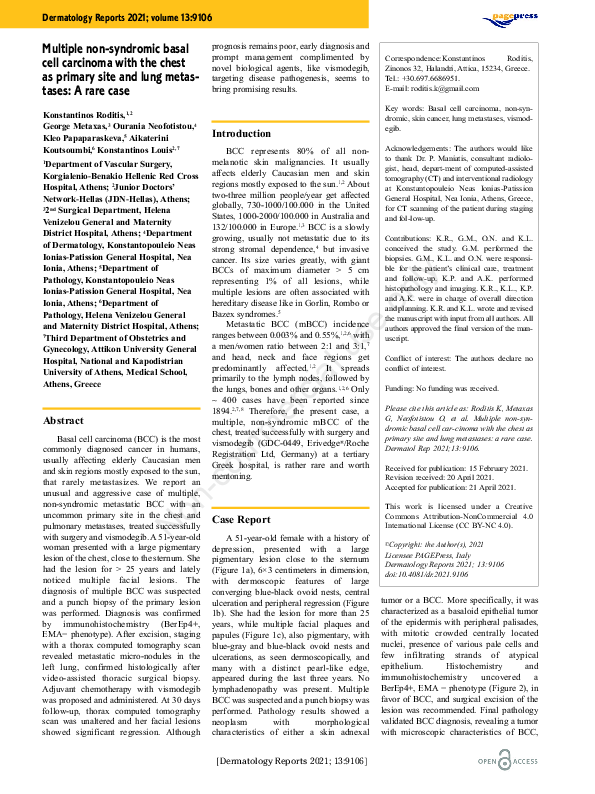
A 51-year-old female with a history of
depression, presented with a large
pigmentary lesion close to the sternum
(Figure 1a), 6×3 centimeters in dimension,
with dermoscopic features of large
converging blue-black ovoid nests, central
ulceration and peripheral regression (Figure
1b). She had the lesion for more than 25
years, while multiple facial plaques and
papules (Figure 1c), also pigmentary, with
blue-gray and blue-black ovoid nests and
ulcerations, as seen dermoscopically, and
many with a distinct pearl-like edge,
appeared during the last three years. No
lymphadenopathy was present. Multiple
BCC was suspected and a punch biopsy was
performed. Pathology results showed a
neoplasm
with
morphological
characteristics of either a skin adnexal
[Dermatology Reports 2021; 13:9106]
ly
Contributions: K.R., G.M., O.N. and K.L.
conceived the study. G.M. performed the
biopsies. G.M., K.L. and O.N. were responsible for the patient’s clinical care, treatment
and follow-up. K.P. and A.K. performed
histopathology and imaging. K.R., K.L., K.P.
and A.K. were in charge of overall direction
and planning. K.R. and K.L. wrote and revised
the manuscript with input from all authors. All
authors approved the final version of the manuscript.
on
e
BCC represents 80% of all nonmelanotic skin malignancies. It usually
affects elderly Caucasian men and skin
regions mostly exposed to the sun.1,2 About
two-three million people/year get affected
globally, 730-1000/100.000 in the United
States, 1000-2000/100.000 in Australia and
132/100.000 in Europe.1,3 BCC is a slowly
growing, usually not metastatic due to its
strong stromal dependence,4 but invasive
cancer. Its size varies greatly, with giant
BCCs of maximum diameter > 5 cm
representing 1% of all lesions, while
multiple lesions are often associated with
hereditary disease like in Gorlin, Rombo or
Bazex syndromes.5
Metastatic BCC (mBCC) incidence
ranges between 0.003% and 0.55%,1,2,6 with
a men/women ratio between 2:1 and 3:1,7
and head, neck and face regions get
predominantly affected.1,2 It spreads
primarily to the lymph nodes, followed by
the lungs, bones and other organs.1,2,6 Only
~ 400 cases have been reported since
1894.2,7,8 Therefore, the present case, a
multiple, non-syndromic mBCC of the
chest, treated successfully with surgery and
vismodegib (GDC-0449, Erivedge®/Roche
Registration Ltd, Germany) at a tertiary
Greek hospital, is rather rare and worth
mentoning.
er
Department of Vascular Surgery,
Korgialenio-Benakio Hellenic Red Cross
Hospital, Athens; 2Junior Doctors’
Network-Hellas (JDN-Hellas), Athens;
3 nd
2 Surgical Department, Helena
Venizelou General and Maternity
District Hospital, Athens; 4Department
of Dermatology, Konstantopouleio Neas
Ionias-Patission General Hospital, Nea
Ionia, Athens; 5Department of
Pathology, Konstantopouleio Neas
Ionias-Patission General Hospital, Nea
Ionia, Athens; 6Department of
Pathology, Helena Venizelou General
and Maternity District Hospital, Athens;
7Third Department of Obstetrics and
Gynecology, Attikon University General
Hospital, National and Kapodistrian
University of Athens, Medical School,
Athens, Greece
Introduction
us
1
Correspondence:Konstantinos
Roditis,
Zinonos 32, Halandri, Attica, 15234, Greece.
Tel.: +30.697.6686951.
E-mail: roditis.k@gmail.com
Key words: Basal cell carcinoma, non-syndromic, skin cancer, lung metastases, vismodegib.
al
Konstantinos Roditis,1,2
George Metaxas,3 Ourania Neofotistou,4
Kleo Papaparaskeva,5 Aikaterini
Koutsoumbi,6 Konstantinos Louis2,7
prognosis remains poor, early diagnosis and
prompt management complimented by
novel biological agents, like vismodegib,
targeting disease pathogenesis, seems to
bring promising results.
ci
Multiple non-syndromic basal
cell carcinoma with the chest
as primary site and lung metastases: A rare case
Conflict of interest: The authors declare no
conflict of interest.
Funding: No funding was received.
Please cite this article as: Roditis K, Metaxas
G, Neofotistou O, et al. Multiple non-syndromic basal cell car-cinoma with the chest as
primary site and lung metastases: a rare case.
Dermatol Rep 2021;13:9106.
Received for publication: 15 February 2021.
Revision received: 20 April 2021.
Accepted for publication: 21 April 2021.
This work is licensed under a Creative
Commons Attribution-NonCommercial 4.0
International License (CC BY-NC 4.0).
©Copyright: the Author(s), 2021
Licensee PAGEPress, Italy
Dermatology Reports 2021; 13:9106
doi:10.4081/dr.2021.9106
tumor or a BCC. More specifically, it was
characterized as a basaloid epithelial tumor
of the epidermis with peripheral palisades,
with mitotic crowded centrally located
nuclei, presence of various pale cells and
few infiltrating strands of atypical
epithelium.
Histochemistry
and
immunohistochemistry
uncovered
a
BerEp4+, EMA − phenotype (Figure 2), in
favor of BCC, and surgical excision of the
lesion was recommended. Final pathology
validated BCC diagnosis, revealing a tumor
with microscopic characteristics of BCC,
�Case Report
Figure 1. a) Unusual location of the primary lesion in the chest, close to the sternum, b)
close view (5×) of the primary lesion, sized 6×3 cm with central ulceration and peripheral
regression, c) multiple facial pearl-like edged lesions with partial ulceration.
on
ly
with various sites of focal ulceration, partial
infiltration of the subcutaneous nerves and
vessels, and minor focal metaplasia to
metatypical basal cell carcinoma, with
negative excision margins. A thorax
computer-assisted tomography scan (CT)
for staging revealed a 7-mm nodule (Figure
3a) and several “ground glass”-like micronodules in the left lung, representing
possible distant metastases. A videoassisted thoracic surgical (VATS) biopsy of
the 7-mm nodule was performed followed
by histological examination of the
specimen, revealing nests of basaloid
epithelial cells with peripheral palisading,
thus confirming its metastatic nature. Due
to disease extent, adjuvant treatment with
vismodegib was initiated at 150mg/day and
she was discharged. An unaltered CT-thorax
(Figure 3b) and regression of her facial
lesions were observed at 30-days follow-up.
us
al
ci
er
m
N
on
-c
om
The usual BCC case is a male, middleaged patient, with a singular small (<5 cm)
non-metastatic lesion located in the head or
neck. Here, we present a multiple mBCC in
a female patient, with a giant (6×3 cm)
primary lesion located in the chest and left
lung metastases, in other words, an unusual
and aggressive form of this common
disease.
Known risk factors, namely light skin
color (Fitzpatrick skin types I and II),
smoking history and occasional exposure to
ultraviolet-A light (UVA), were present,
while exposure to arsenic, ionizing
radiation or dry ice, immunosupression,
albinism, keratoacanthoma or xeroderma
pigmentosum were absent. Hereditary
multiple BCC was excluded in our case due
to
negative
family
history
of
genodermatoses.1,3,5
Although the fourth, fifth and sixth
decades are considered the mean or median
age of presentation of the primary lesion,2,7
our patient developed her first BCC at age
26. Nevertheless, it was left untreated for 25
years and not only it metastasized, but she
also developed multiple primary lesions, as
expected by described high 1-year and 5year cumulative risks to develop secondary
lesions.5,6,9
Metastatic BCC occurs two times more
often in males than females with the most
common site of metastases being lymph
nodes, followed by the lungs. Presentation
age, the site and size of primary lesion,
perineural and perivascular spread, duration
and recurrence of disease, incomplete
resection or positive margins, multiple
e
Discussion
Figure 2. Comparison BerEp4(﹢) immunohistochemistry stain (200×) (a) with EMA(−)
(200×) (b) and H&E (400×) (c) stains of the primary tumor punch biopsy.
[Dermatology Reports 2021; 13:9106]
[page 71]
�Case Report
doctor-patient relationship regarding
communication of knowledge on the
primary BCC disease and its metastatic
potential, can indeed play a crucial role in
increasing favorable outcomes. Precise
excision of the primary tumor and adjuvant
targeted therapy with Hedgehog inhibitors,
can bring promising results in aggressive
cases of mBCC and must be encouraged.
References
us
e
on
ly
1. Rubin AI, Chen EH, Ratner D. Basalcell carcinoma. N Engl J Med
2005;353: 2262-9.
2. Piva de Freitas P, Senna CG, Tabai M,
et al. Metastatic basal cell carcinoma: a
rare manifestation of a common disease. Case Rep Med 2017;2017:
8929745.
3. Wu S, Han J, Li WQ, et al. Basal-cell
carcinoma incidence and associated risk
factors in U.S. women and men. Am J
Epidemiol 2013;178:890-7.
4. Robinson JK. Risk of developing another basal cell carcinoma: a 5-year
prospective study. Cancer 1987;60:11820.
5. Kim DH, Ko HS, Jun YJ.
Nonsyndromic Multiple Basal Cell
Carcinomas. Arch Craniofac Surg
2017;18:191-6.
6. Robinson JK, Dahiya M. Basal cell carcinoma with pulmonary and lymph
node metastasis causing death. Arch
Dermatol 2003;139:643-8.
7. Wysong A, Aasi SZ, Tang JY. Update
on metastatic basal cell carcinoma: a
summary of published cases from 1981
through 2011. JAMA Dermatol
2013;149:615-6.
8. Lattes R, Kessler RW. Metastasizing
basal-cell epithelioma of the skin;
report of two cases. Cancer 1951;4:86678.
9. Marcil I, Stern RS. Risk of developing a
subsequent nonmelanoma skin cancer
in patients with a history of nonmelanoma skin cancer: a critical review
of the literature and meta-analysis. Arch
Dermatol 2000;136:1524-30.
10. Meiss F, Andrlová H, Zeiser R.
Vismodegib. Recent Results Cancer
Res 2018;211:125-39.
om
m
er
indicated for metastatic disease.10 Although
nausea/loss of appetite and vomiting,
diarrhea or constipation, weight and hair
loss, muscle spasms and joint pain have
been described among others as common
side-effects, response rates can reach 37%,
thus proving their therapeutic potential
against mBCC.2 Our patient received this
novel therapy and showed positive primary
results.
N
on
-c
tumors and aggressive histological types
may predict metastasis.1-3 Tumors greater
than 3 cm in diameter have a 2% incidence
of metastasis and/or death.2,6 Prognosis is
poor,1,2,6,7 with a longer average survival of
three-and-a-half to seven years, when
lymphatic spread is involved,2 in contrast
with an average of eight months to two
years that accompanies hematogenous
metastases.1,2,4,7
Surgery and radiotherapy combined or
not with targeted therapy are the most
common therapeutic options used.2,5
Aberrant activation of the Hh pathway has
been associated with pathogenesis of
sporadic, non-syndromic BCCs as well as in
hereditary syndromes with multiple
BCCs.1,5 Small molecule antagonists of the
Hedgehog pathway, like vismodegib and
sonidegib, can both be used as adjuvant
targeted therapy in locally advanced
disease, however, only vismodegib is
ci
al
Figure 3. CT-thorax scan images of the metastatic 7 mm left lung nodule: (a) single arrow
- at presentation, (b) double arrow - at 30-days follow-up.
[page 72]
Conclusions
In conclusion, the earlier the
identification of this potentially curable
form of cancer, the better the therapeutic
results and the least the morbidity
complications.
Comprehensive
sensitization of the population and
awareness raising by all relevant
physicians, as well as fostering a healthy
[Dermatology Reports 2021; 13:9106]
�
 Konstantinos Roditis