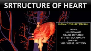
Location via proxy:   [ UP ]  
[Report a bug]   [Manage cookies]                
SlideShare a Scribd company logo
SRTRUCTURE OF HEART
Human Circulatory
System
Human circulatory system,
also called the blood
vascular system consists of
a muscular chambered
heart, a network of closed
branching blood vessels
and blood, the fluid which
is circulated.
The Heart
• Heart is a muscular organ that
pumps blood received from the
veins into the arteries, thereby
maintaining the flow of blood
through the entire circulatory
system.
• Mesodermally derived organ.
• Situated in the thoracic cavity, in
between the two lungs, slightly
tilted to the left.
• Size of a clenched fist.
The Pericardium
• It is protected by a
double walled
membranous bag,
pericardium, enclosing
the pericardial fluid.
• Function - to restrict
excessive movements of
the heart as a whole and
serve as a lubricated
container in which
different parts of heart
can contract.
R Removal of a section of pericardium
Heart enclosed in pericardium

Recommended for you

Cardiac cycle
Cardiac cycleCardiac cycle
Cardiac cycle

The cardiac cycle refers to the repeating sequence of events that occur with each heartbeat, from the beginning of one heartbeat to the beginning of the next. The cardiac cycle involves both atrial and ventricular contraction (systole) followed by relaxation (diastole). There are three main stages: 1) atrial systole, where the atria contract; 2) ventricular systole, where the ventricles contract; and 3) complete cardiac diastole, where both the atria and ventricles relax. The cycle allows for blood to flow through the heart and into the arteries with each heartbeat.

SINOATRIAL NODE
SINOATRIAL NODESINOATRIAL NODE
SINOATRIAL NODE

The sinoatrial node, located in the wall of the right atrium, is the heart's natural pacemaker. It generates electrical impulses that pass through the heart and cause it to contract. The sinoatrial node initiates action potentials that travel through the conduction system to the atrioventricular node and then to the ventricles. As the primary pacemaker, the sinoatrial node controls the heart rate by regulating the firing of action potentials. Dysfunction or ischemia of the sinoatrial node can lead to irregular heart rhythms.

sa node 01
Cardiac cycle
Cardiac cycleCardiac cycle
Cardiac cycle

The cardiac cycle describes the sequence of events that occur with each heartbeat. It consists of systole, when the heart contracts and pumps blood, and diastole, when the heart relaxes and fills with blood. The cardiac cycle includes atrial and ventricular events. Atrial systole forces a small amount of blood into the ventricles, while atrial diastole allows the atria to fill with blood. Ventricular events include isovolumetric contraction, ejection of blood, isovolumetric relaxation, and rapid and slow filling as the ventricles refill with blood. The pressures and volumes in the heart chambers change throughout the cardiac cycle.

Heart Walls
1. Epicardium – visceral layer of the serous pericardium. outer protective
layer.
2. Myocardium – cardiac muscle layer forming the bulk of the heart. It is
the middle layer of the heart. Fibrous skeleton of the heart –
crisscrossing, interlacing layer of connective tissue.
3. Endocardium – endothelial layer of the inner myocardial surface. It is
the deepest layer of the heart. It has the smooth lining to reduce
friction of blood flow.
The Heart Chambers
Our heart has 4 chambers 
1. Two relatively small upper chambers called atria
2. Two larger lower chambers called ventricles
• The right atrium receives blood from the body. This
blood is low in oxygen. This is the blood from the
veins.
• The right ventricle pumps the blood from the right
atrium into the lungs to pick up oxygen and remove
carbon dioxide.
• The left atrium receives blood from the lungs. This
blood is rich in oxygen.
• The left ventricle pumps the blood from the left
atrium out to the body, supplying all organs with
oxygen-rich blood.
Blood Vessels Of Heart
These bring blood to the lungs, where
oxygen enters the bloodstream, and then
to the body:
• The inferior and superior vena cava bring
deoxygenated blood from the body into
the right atrium.
• The pulmonary artery channels oxygen-
poor blood from the right ventricle into the
lungs, where oxygen enters the
bloodstream.
• The pulmonary veins bring oxygen-
rich blood to the left atrium.
• The aorta channels oxygen-rich blood to
the body from the left ventricle.
Coronary Circulation
Coronary circulation is the circulation of
blood in the blood vessels that supply
the heart muscle (myocardium).
Coronary arteries supply oxygenated
blood to the heart muscle, and cardiac
veins drain away the blood once it has
been deoxygenated.
• Two coronary arteries arise from
the aorta just beyond the semilunar
valves; supply blood into the musculature
of the heart.
• Deoxygenated blood is returned to the
chambers of the heart via coronary veins;
most of these converge to form the
coronary venous sinus, which drains into
the right atrium.

Recommended for you

Cardiac output 1
Cardiac output 1Cardiac output 1
Cardiac output 1

This document discusses cardiac output and its measurement. It defines cardiac output as the amount of blood ejected by each ventricle per minute, which is calculated as stroke volume multiplied by heart rate. It describes several methods to measure cardiac output, including the indicator dye dilution method, thermodilution, and measurement of inhaled inert gases. It discusses factors that can cause cardiac output to vary, such as age, sex, environmental temperature, exercise, and various pathological conditions.

dr nilesh kate cardiac output
Heart rate by pandian m
Heart rate by pandian mHeart rate by pandian m
Heart rate by pandian m

HEART RATE REGULATION OF HEART RATE VASOMOTOR CENTER – CARDIAC CENTER MOTOR (EFFERENT) NERVE FIBERS TO HEART FACTORS AFFECTING VASOMOTOR CENTER for all medical & health care students

#The heart as a pump and function of the heart valves
#The heart as a pump and function of the heart valves#The heart as a pump and function of the heart valves
#The heart as a pump and function of the heart valves

This document discusses the physiology of the heart. It begins by describing the different types of cardiac muscle and how cardiac muscle cells are interconnected. It then covers the cardiac cycle, including diastole and systole. Action potentials in cardiac muscle are longer than in skeletal muscle due to slow calcium channels. Contraction is triggered by calcium release from the sarcoplasmic reticulum and extracellular fluids. The heart pumps in two stages - the atria prime the ventricles, then the ventricles eject blood. Various waves in pressures, ECG, and sounds are related to the different cardiac cycle events.

The Septa
• A thin, muscular wall called the
interatrial septum separates the
right and the left atria.
• Whereas a thick-walled, the
inter-ventricular septum,
separates the left and the right
ventricles.
• The atrium and the ventricle of
the same side are also separated
by a thick fibrous tissue called the
atrio-ventricular septum.
Atrio-ventricular septum
Interatrial Septum
Interventricular septum
Heart Valves
Each of the septa are provided with an
opening through which the two chambers
of the same side are connected.
• The opening between the right atrium &
the right ventricle is guarded by a valve
formed of 3 muscular flaps or cusps, the
tricuspid valve.
• A bicuspid/mitral valve guards the opening
b/w the left atrium & the left ventricle.
• The openings of the right and the left
ventricles into the pulmonary artery and
the aorta are provided with the semilunar
valves.
The valves in the heart allows the flow of
blood only in one direction, i.e., from the
atria to the ventricles and from the
ventricles to the pulmonary artery or aorta.
These valves prevent any backward flow.
Myogenic Nature Of Heart
A myogenic heart is capable of
generating a cardiac contraction
independent of nervous input. In a
myogenic heart contraction is initiated
by the myocyte cell itself instead of an
outside occurrence or stimulus such as
nerve innervation.
Reason-
• A specialized cardiac musculature called
the nodal musculature is distributed in
the heart.
The Nodal Tissue
1. A patch of this tissue is present in the right
upper corner of the right atrium-sino-atrial
node (SAN).
2. Another mass of this tissue is seen in the
lower left corner of the right atrium close
to the atrio-ventricular septum-atrio-
ventricular node (AVN).
3. Bundle of nodal fibers, atrioventricular
bundle (AV bundle) continues from the
AVN, passes through the atrio-ventricular
septa to emerge on the top of the
interventricular septum and immediately
divides into a right and left AV bundle.
4. These branches give rise to minute fibers
throughout the ventricular musculature of
the respective sides and are called purkinje
fibers.

Recommended for you

Heart anatomy and functions
Heart anatomy and functionsHeart anatomy and functions
Heart anatomy and functions

DESCRIPTION ABOUT HEART ANATOMICAL STRUCTURES, FUNCTIONS OF HEART, PARTS OF HEART THEIR BASIC PATHWAYS FOR BLOOD CIRCULATIONS, TYPES OF PATHWAYS ALSO, ANATOMICAL PICTURE OF HEART.

cardiovascular physiology
 cardiovascular physiology cardiovascular physiology
cardiovascular physiology

The document provides an overview of cardiovascular physiology, including: - The cardiovascular system functions to circulate blood throughout the body, transporting oxygen, nutrients, hormones, and removing waste. - The heart is the central organ that pumps blood through two main circulations - pulmonary circulation to the lungs and systemic circulation to the rest of the body. - The functional anatomy of the heart includes four chambers, cardiac muscle tissue, valves that ensure one-way blood flow, and a conducting system that coordinates contractions.

Cardiac output
Cardiac outputCardiac output
Cardiac output

Cardiac output refers to the volume of blood pumped by each ventricle per minute. It is determined by stroke volume, heart rate, preload, afterload, and peripheral resistance. Normal cardiac output is approximately 5 liters per minute. It can increase up to 600% in athletes due to enhanced cardiac reserve. Factors such as venous return, force of contraction, heart rate, and peripheral resistance maintain cardiac output within the body.

• The nodal musculature has the ability to generate action potentials
without any external stimuli, i.e., it is auto excitable.
• However, the number of action potentials that could be generated in
a minute vary at different parts of the nodal system.
• The SAN can generate the maximum number of action potentials, i.e.,
70-75 min–1 , and is responsible for initiating and maintaining the
rhythmic contractile activity of the heart.
• Therefore, SAN is called the pacemaker.
• Our heart normally beats 70-75 times in a minute (average 72 beats
min–1).
Actional Potential Generation
Thank you

More Related Content

What's hot

blood supply of heart
blood supply of heartblood supply of heart
Conduction System of the Heart
Conduction System of the HeartConduction System of the Heart
Conduction System of the Heart
DR. CARLOS Azañero
 
Blood vessels of the heart
Blood vessels of the heartBlood vessels of the heart
Blood vessels of the heart
jithahari
 
Cardiac cycle
Cardiac cycleCardiac cycle
Cardiac cycle
Kashikant Yadav
 
SINOATRIAL NODE
SINOATRIAL NODESINOATRIAL NODE
SINOATRIAL NODE
Bidhan Sarkar
 
Cardiac cycle
Cardiac cycleCardiac cycle
Cardiac cycle
mariaidrees3
 
Cardiac output 1
Cardiac output 1Cardiac output 1
Cardiac output 1
Dr Nilesh Kate
 
Heart rate by pandian m
Heart rate by pandian mHeart rate by pandian m
Heart rate by pandian m
Physiology Dept
 
#The heart as a pump and function of the heart valves
#The heart as a pump and function of the heart valves#The heart as a pump and function of the heart valves
#The heart as a pump and function of the heart valves
Lubna Abu Alrub,DDS
 
Heart anatomy and functions
Heart anatomy and functionsHeart anatomy and functions
Heart anatomy and functions
MUSKANKr
 
cardiovascular physiology
 cardiovascular physiology cardiovascular physiology
cardiovascular physiology
aliagr
 
Cardiac output
Cardiac outputCardiac output
Cardiac output
mariaidrees3
 
Regional circulations
Regional circulationsRegional circulations
Regional circulations
vajira54
 
Pericardium
PericardiumPericardium
Pericardium
Nikhil Vaishnav
 
Location & structure of the heart
Location & structure  of the heartLocation & structure  of the heart
Location & structure of the heart
Vyramuthu Atputhan
 
Valves of heart
Valves of heartValves of heart
Valves of heart
Nikhil Vaishnav
 
Heart anatomy
Heart anatomyHeart anatomy
Heart anatomy
RobbinsHobbin
 
The coronary circulation
The coronary circulationThe coronary circulation
The coronary circulation
Idris Siddiqui
 
Cardiovascular system (blood pressure, hypertension)
Cardiovascular system (blood pressure, hypertension) Cardiovascular system (blood pressure, hypertension)
Cardiovascular system (blood pressure, hypertension)
Pharmacy Universe
 
Bronchial tree
Bronchial treeBronchial tree
Bronchial tree
Medical Students
 

What's hot (20)

blood supply of heart
blood supply of heartblood supply of heart
blood supply of heart
 
Conduction System of the Heart
Conduction System of the HeartConduction System of the Heart
Conduction System of the Heart
 
Blood vessels of the heart
Blood vessels of the heartBlood vessels of the heart
Blood vessels of the heart
 
Cardiac cycle
Cardiac cycleCardiac cycle
Cardiac cycle
 
SINOATRIAL NODE
SINOATRIAL NODESINOATRIAL NODE
SINOATRIAL NODE
 
Cardiac cycle
Cardiac cycleCardiac cycle
Cardiac cycle
 
Cardiac output 1
Cardiac output 1Cardiac output 1
Cardiac output 1
 
Heart rate by pandian m
Heart rate by pandian mHeart rate by pandian m
Heart rate by pandian m
 
#The heart as a pump and function of the heart valves
#The heart as a pump and function of the heart valves#The heart as a pump and function of the heart valves
#The heart as a pump and function of the heart valves
 
Heart anatomy and functions
Heart anatomy and functionsHeart anatomy and functions
Heart anatomy and functions
 
cardiovascular physiology
 cardiovascular physiology cardiovascular physiology
cardiovascular physiology
 
Cardiac output
Cardiac outputCardiac output
Cardiac output
 
Regional circulations
Regional circulationsRegional circulations
Regional circulations
 
Pericardium
PericardiumPericardium
Pericardium
 
Location & structure of the heart
Location & structure  of the heartLocation & structure  of the heart
Location & structure of the heart
 
Valves of heart
Valves of heartValves of heart
Valves of heart
 
Heart anatomy
Heart anatomyHeart anatomy
Heart anatomy
 
The coronary circulation
The coronary circulationThe coronary circulation
The coronary circulation
 
Cardiovascular system (blood pressure, hypertension)
Cardiovascular system (blood pressure, hypertension) Cardiovascular system (blood pressure, hypertension)
Cardiovascular system (blood pressure, hypertension)
 
Bronchial tree
Bronchial treeBronchial tree
Bronchial tree
 

Similar to Structure Of Heart

UNIT 4-WPS Office.pptx
UNIT 4-WPS Office.pptxUNIT 4-WPS Office.pptx
UNIT 4-WPS Office.pptx
DNYANESHWARBIRADAR1
 
7. cardiovascular system
7. cardiovascular system7. cardiovascular system
7. cardiovascular system
Yogeshwary Bhongade
 
The Circulatory System.pptx
The Circulatory System.pptxThe Circulatory System.pptx
The Circulatory System.pptx
NehaPandey199
 
Transport in animals
Transport in animalsTransport in animals
Transport in animals
Sian Ferguson
 
CARDIOVASCULAR SYSTEM.pptx
CARDIOVASCULAR SYSTEM.pptxCARDIOVASCULAR SYSTEM.pptx
CARDIOVASCULAR SYSTEM.pptx
Sweety gupta
 
Anatomy of Human Heart along with internal and external features.
Anatomy of Human Heart along with internal and external features.Anatomy of Human Heart along with internal and external features.
Anatomy of Human Heart along with internal and external features.
ssuser7bf0a2
 
Cardiovascular System unit VIII - Copy Std.pptx
Cardiovascular System unit VIII - Copy Std.pptxCardiovascular System unit VIII - Copy Std.pptx
Cardiovascular physiology for university students
Cardiovascular physiology for university studentsCardiovascular physiology for university students
Cardiovascular physiology for university students
ItsOnyii
 
CARDIO VASCULAR SYSTEM THE HEART
CARDIO VASCULAR SYSTEM THE HEARTCARDIO VASCULAR SYSTEM THE HEART
CARDIO VASCULAR SYSTEM THE HEART
PARUL UNIVERSITY
 
CARDIOVASCULAR SYSTEM
CARDIOVASCULAR SYSTEM CARDIOVASCULAR SYSTEM
CARDIOVASCULAR SYSTEM
M M J G COLLEGE OF PHARMACY
 
3-180318193312.pdf
3-180318193312.pdf3-180318193312.pdf
3-180318193312.pdf
snithiyuvarajayuvara
 
3. internal structure of the heart
3. internal structure of the heart3. internal structure of the heart
3. internal structure of the heart
Dr. Mohammad Mahmoud
 
Cardiovascular Anatomy.pptx
Cardiovascular Anatomy.pptxCardiovascular Anatomy.pptx
Cardiovascular Anatomy.pptx
xamdathisbad
 
Cardiovascular Anatomy.pptx
Cardiovascular Anatomy.pptxCardiovascular Anatomy.pptx
Cardiovascular Anatomy.pptx
xamdathisbad
 
Cardiovascular system by Yogesh patel
Cardiovascular system by Yogesh patelCardiovascular system by Yogesh patel
Cardiovascular system by Yogesh patel
Prabhat Kumar
 
anatomy and physiology of cardiovascular 1 (1).pptx
anatomy and physiology of cardiovascular 1 (1).pptxanatomy and physiology of cardiovascular 1 (1).pptx
anatomy and physiology of cardiovascular 1 (1).pptx
EricsonKiprono
 
Johny's A&P Structure and Function of Heart
Johny's A&P Structure and Function of HeartJohny's A&P Structure and Function of Heart
Johny's A&P Structure and Function of Heart
Johny Kutty Joseph
 
heart anatomy pdf.pdf
heart anatomy pdf.pdfheart anatomy pdf.pdf
heart anatomy pdf.pdf
Pooja Rani
 
Structure of heart
Structure of heartStructure of heart
Structure of heart
sukh randhawa
 
Human heart
Human heartHuman heart
Human heart
Hussein Al-tameemi
 

Similar to Structure Of Heart (20)

UNIT 4-WPS Office.pptx
UNIT 4-WPS Office.pptxUNIT 4-WPS Office.pptx
UNIT 4-WPS Office.pptx
 
7. cardiovascular system
7. cardiovascular system7. cardiovascular system
7. cardiovascular system
 
The Circulatory System.pptx
The Circulatory System.pptxThe Circulatory System.pptx
The Circulatory System.pptx
 
Transport in animals
Transport in animalsTransport in animals
Transport in animals
 
CARDIOVASCULAR SYSTEM.pptx
CARDIOVASCULAR SYSTEM.pptxCARDIOVASCULAR SYSTEM.pptx
CARDIOVASCULAR SYSTEM.pptx
 
Anatomy of Human Heart along with internal and external features.
Anatomy of Human Heart along with internal and external features.Anatomy of Human Heart along with internal and external features.
Anatomy of Human Heart along with internal and external features.
 
Cardiovascular System unit VIII - Copy Std.pptx
Cardiovascular System unit VIII - Copy Std.pptxCardiovascular System unit VIII - Copy Std.pptx
Cardiovascular System unit VIII - Copy Std.pptx
 
Cardiovascular physiology for university students
Cardiovascular physiology for university studentsCardiovascular physiology for university students
Cardiovascular physiology for university students
 
CARDIO VASCULAR SYSTEM THE HEART
CARDIO VASCULAR SYSTEM THE HEARTCARDIO VASCULAR SYSTEM THE HEART
CARDIO VASCULAR SYSTEM THE HEART
 
CARDIOVASCULAR SYSTEM
CARDIOVASCULAR SYSTEM CARDIOVASCULAR SYSTEM
CARDIOVASCULAR SYSTEM
 
3-180318193312.pdf
3-180318193312.pdf3-180318193312.pdf
3-180318193312.pdf
 
3. internal structure of the heart
3. internal structure of the heart3. internal structure of the heart
3. internal structure of the heart
 
Cardiovascular Anatomy.pptx
Cardiovascular Anatomy.pptxCardiovascular Anatomy.pptx
Cardiovascular Anatomy.pptx
 
Cardiovascular Anatomy.pptx
Cardiovascular Anatomy.pptxCardiovascular Anatomy.pptx
Cardiovascular Anatomy.pptx
 
Cardiovascular system by Yogesh patel
Cardiovascular system by Yogesh patelCardiovascular system by Yogesh patel
Cardiovascular system by Yogesh patel
 
anatomy and physiology of cardiovascular 1 (1).pptx
anatomy and physiology of cardiovascular 1 (1).pptxanatomy and physiology of cardiovascular 1 (1).pptx
anatomy and physiology of cardiovascular 1 (1).pptx
 
Johny's A&P Structure and Function of Heart
Johny's A&P Structure and Function of HeartJohny's A&P Structure and Function of Heart
Johny's A&P Structure and Function of Heart
 
heart anatomy pdf.pdf
heart anatomy pdf.pdfheart anatomy pdf.pdf
heart anatomy pdf.pdf
 
Structure of heart
Structure of heartStructure of heart
Structure of heart
 
Human heart
Human heartHuman heart
Human heart
 

Recently uploaded

AI Risk Management: ISO/IEC 42001, the EU AI Act, and ISO/IEC 23894
AI Risk Management: ISO/IEC 42001, the EU AI Act, and ISO/IEC 23894AI Risk Management: ISO/IEC 42001, the EU AI Act, and ISO/IEC 23894
AI Risk Management: ISO/IEC 42001, the EU AI Act, and ISO/IEC 23894
PECB
 
Beyond the Advance Presentation for By the Book 9
Beyond the Advance Presentation for By the Book 9Beyond the Advance Presentation for By the Book 9
Beyond the Advance Presentation for By the Book 9
John Rodzvilla
 
Beginner's Guide to Bypassing Falco Container Runtime Security in Kubernetes ...
Beginner's Guide to Bypassing Falco Container Runtime Security in Kubernetes ...Beginner's Guide to Bypassing Falco Container Runtime Security in Kubernetes ...
Beginner's Guide to Bypassing Falco Container Runtime Security in Kubernetes ...
anjaliinfosec
 
Delegation Inheritance in Odoo 17 and Its Use Cases
Delegation Inheritance in Odoo 17 and Its Use CasesDelegation Inheritance in Odoo 17 and Its Use Cases
Delegation Inheritance in Odoo 17 and Its Use Cases
Celine George
 
Front Desk Management in the Odoo 17 ERP
Front Desk  Management in the Odoo 17 ERPFront Desk  Management in the Odoo 17 ERP
Front Desk Management in the Odoo 17 ERP
Celine George
 
Final ebook Keeping the Memory @live.pdf
Final ebook Keeping the Memory @live.pdfFinal ebook Keeping the Memory @live.pdf
Final ebook Keeping the Memory @live.pdf
Zuzana Mészárosová
 
ENGLISH-7-CURRICULUM MAP- MATATAG CURRICULUM
ENGLISH-7-CURRICULUM MAP- MATATAG CURRICULUMENGLISH-7-CURRICULUM MAP- MATATAG CURRICULUM
ENGLISH-7-CURRICULUM MAP- MATATAG CURRICULUM
HappieMontevirgenCas
 
The basics of sentences session 9pptx.pptx
The basics of sentences session 9pptx.pptxThe basics of sentences session 9pptx.pptx
The basics of sentences session 9pptx.pptx
heathfieldcps1
 
Capitol Doctoral Presentation -June 2024v2.pptx
Capitol Doctoral Presentation -June 2024v2.pptxCapitol Doctoral Presentation -June 2024v2.pptx
Capitol Doctoral Presentation -June 2024v2.pptx
CapitolTechU
 
Views in Odoo - Advanced Views - Pivot View in Odoo 17
Views in Odoo - Advanced Views - Pivot View in Odoo 17Views in Odoo - Advanced Views - Pivot View in Odoo 17
Views in Odoo - Advanced Views - Pivot View in Odoo 17
Celine George
 
The Jewish Trinity : Sabbath,Shekinah and Sanctuary 4.pdf
The Jewish Trinity : Sabbath,Shekinah and Sanctuary 4.pdfThe Jewish Trinity : Sabbath,Shekinah and Sanctuary 4.pdf
The Jewish Trinity : Sabbath,Shekinah and Sanctuary 4.pdf
JackieSparrow3
 
Understanding and Interpreting Teachers’ TPACK for Teaching Multimodalities i...
Understanding and Interpreting Teachers’ TPACK for Teaching Multimodalities i...Understanding and Interpreting Teachers’ TPACK for Teaching Multimodalities i...
Understanding and Interpreting Teachers’ TPACK for Teaching Multimodalities i...
Neny Isharyanti
 
2024 KWL Back 2 School Summer Conference
2024 KWL Back 2 School Summer Conference2024 KWL Back 2 School Summer Conference
2024 KWL Back 2 School Summer Conference
KlettWorldLanguages
 
How to Show Sample Data in Tree and Kanban View in Odoo 17
How to Show Sample Data in Tree and Kanban View in Odoo 17How to Show Sample Data in Tree and Kanban View in Odoo 17
How to Show Sample Data in Tree and Kanban View in Odoo 17
Celine George
 
Split Shifts From Gantt View in the Odoo 17
Split Shifts From Gantt View in the  Odoo 17Split Shifts From Gantt View in the  Odoo 17
Split Shifts From Gantt View in the Odoo 17
Celine George
 
How to Store Data on the Odoo 17 Website
How to Store Data on the Odoo 17 WebsiteHow to Store Data on the Odoo 17 Website
How to Store Data on the Odoo 17 Website
Celine George
 
ARCHITECTURAL PATTERNS IN HISTOPATHOLOGY pdf- [Autosaved].pdf
ARCHITECTURAL PATTERNS IN HISTOPATHOLOGY  pdf-  [Autosaved].pdfARCHITECTURAL PATTERNS IN HISTOPATHOLOGY  pdf-  [Autosaved].pdf
ARCHITECTURAL PATTERNS IN HISTOPATHOLOGY pdf- [Autosaved].pdf
DharmarajPawar
 
How to Configure Time Off Types in Odoo 17
How to Configure Time Off Types in Odoo 17How to Configure Time Off Types in Odoo 17
How to Configure Time Off Types in Odoo 17
Celine George
 
Ardra Nakshatra (आर्द्रा): Understanding its Effects and Remedies
Ardra Nakshatra (आर्द्रा): Understanding its Effects and RemediesArdra Nakshatra (आर्द्रा): Understanding its Effects and Remedies
Ardra Nakshatra (आर्द्रा): Understanding its Effects and Remedies
Astro Pathshala
 
(T.L.E.) Agriculture: Essentials of Gardening
(T.L.E.) Agriculture: Essentials of Gardening(T.L.E.) Agriculture: Essentials of Gardening
(T.L.E.) Agriculture: Essentials of Gardening
MJDuyan
 

Recently uploaded (20)

AI Risk Management: ISO/IEC 42001, the EU AI Act, and ISO/IEC 23894
AI Risk Management: ISO/IEC 42001, the EU AI Act, and ISO/IEC 23894AI Risk Management: ISO/IEC 42001, the EU AI Act, and ISO/IEC 23894
AI Risk Management: ISO/IEC 42001, the EU AI Act, and ISO/IEC 23894
 
Beyond the Advance Presentation for By the Book 9
Beyond the Advance Presentation for By the Book 9Beyond the Advance Presentation for By the Book 9
Beyond the Advance Presentation for By the Book 9
 
Beginner's Guide to Bypassing Falco Container Runtime Security in Kubernetes ...
Beginner's Guide to Bypassing Falco Container Runtime Security in Kubernetes ...Beginner's Guide to Bypassing Falco Container Runtime Security in Kubernetes ...
Beginner's Guide to Bypassing Falco Container Runtime Security in Kubernetes ...
 
Delegation Inheritance in Odoo 17 and Its Use Cases
Delegation Inheritance in Odoo 17 and Its Use CasesDelegation Inheritance in Odoo 17 and Its Use Cases
Delegation Inheritance in Odoo 17 and Its Use Cases
 
Front Desk Management in the Odoo 17 ERP
Front Desk  Management in the Odoo 17 ERPFront Desk  Management in the Odoo 17 ERP
Front Desk Management in the Odoo 17 ERP
 
Final ebook Keeping the Memory @live.pdf
Final ebook Keeping the Memory @live.pdfFinal ebook Keeping the Memory @live.pdf
Final ebook Keeping the Memory @live.pdf
 
ENGLISH-7-CURRICULUM MAP- MATATAG CURRICULUM
ENGLISH-7-CURRICULUM MAP- MATATAG CURRICULUMENGLISH-7-CURRICULUM MAP- MATATAG CURRICULUM
ENGLISH-7-CURRICULUM MAP- MATATAG CURRICULUM
 
The basics of sentences session 9pptx.pptx
The basics of sentences session 9pptx.pptxThe basics of sentences session 9pptx.pptx
The basics of sentences session 9pptx.pptx
 
Capitol Doctoral Presentation -June 2024v2.pptx
Capitol Doctoral Presentation -June 2024v2.pptxCapitol Doctoral Presentation -June 2024v2.pptx
Capitol Doctoral Presentation -June 2024v2.pptx
 
Views in Odoo - Advanced Views - Pivot View in Odoo 17
Views in Odoo - Advanced Views - Pivot View in Odoo 17Views in Odoo - Advanced Views - Pivot View in Odoo 17
Views in Odoo - Advanced Views - Pivot View in Odoo 17
 
The Jewish Trinity : Sabbath,Shekinah and Sanctuary 4.pdf
The Jewish Trinity : Sabbath,Shekinah and Sanctuary 4.pdfThe Jewish Trinity : Sabbath,Shekinah and Sanctuary 4.pdf
The Jewish Trinity : Sabbath,Shekinah and Sanctuary 4.pdf
 
Understanding and Interpreting Teachers’ TPACK for Teaching Multimodalities i...
Understanding and Interpreting Teachers’ TPACK for Teaching Multimodalities i...Understanding and Interpreting Teachers’ TPACK for Teaching Multimodalities i...
Understanding and Interpreting Teachers’ TPACK for Teaching Multimodalities i...
 
2024 KWL Back 2 School Summer Conference
2024 KWL Back 2 School Summer Conference2024 KWL Back 2 School Summer Conference
2024 KWL Back 2 School Summer Conference
 
How to Show Sample Data in Tree and Kanban View in Odoo 17
How to Show Sample Data in Tree and Kanban View in Odoo 17How to Show Sample Data in Tree and Kanban View in Odoo 17
How to Show Sample Data in Tree and Kanban View in Odoo 17
 
Split Shifts From Gantt View in the Odoo 17
Split Shifts From Gantt View in the  Odoo 17Split Shifts From Gantt View in the  Odoo 17
Split Shifts From Gantt View in the Odoo 17
 
How to Store Data on the Odoo 17 Website
How to Store Data on the Odoo 17 WebsiteHow to Store Data on the Odoo 17 Website
How to Store Data on the Odoo 17 Website
 
ARCHITECTURAL PATTERNS IN HISTOPATHOLOGY pdf- [Autosaved].pdf
ARCHITECTURAL PATTERNS IN HISTOPATHOLOGY  pdf-  [Autosaved].pdfARCHITECTURAL PATTERNS IN HISTOPATHOLOGY  pdf-  [Autosaved].pdf
ARCHITECTURAL PATTERNS IN HISTOPATHOLOGY pdf- [Autosaved].pdf
 
How to Configure Time Off Types in Odoo 17
How to Configure Time Off Types in Odoo 17How to Configure Time Off Types in Odoo 17
How to Configure Time Off Types in Odoo 17
 
Ardra Nakshatra (आर्द्रा): Understanding its Effects and Remedies
Ardra Nakshatra (आर्द्रा): Understanding its Effects and RemediesArdra Nakshatra (आर्द्रा): Understanding its Effects and Remedies
Ardra Nakshatra (आर्द्रा): Understanding its Effects and Remedies
 
(T.L.E.) Agriculture: Essentials of Gardening
(T.L.E.) Agriculture: Essentials of Gardening(T.L.E.) Agriculture: Essentials of Gardening
(T.L.E.) Agriculture: Essentials of Gardening
 

Structure Of Heart

  • 2. Human Circulatory System Human circulatory system, also called the blood vascular system consists of a muscular chambered heart, a network of closed branching blood vessels and blood, the fluid which is circulated.
  • 3. The Heart • Heart is a muscular organ that pumps blood received from the veins into the arteries, thereby maintaining the flow of blood through the entire circulatory system. • Mesodermally derived organ. • Situated in the thoracic cavity, in between the two lungs, slightly tilted to the left. • Size of a clenched fist.
  • 4. The Pericardium • It is protected by a double walled membranous bag, pericardium, enclosing the pericardial fluid. • Function - to restrict excessive movements of the heart as a whole and serve as a lubricated container in which different parts of heart can contract. R Removal of a section of pericardium Heart enclosed in pericardium
  • 5. Heart Walls 1. Epicardium – visceral layer of the serous pericardium. outer protective layer. 2. Myocardium – cardiac muscle layer forming the bulk of the heart. It is the middle layer of the heart. Fibrous skeleton of the heart – crisscrossing, interlacing layer of connective tissue. 3. Endocardium – endothelial layer of the inner myocardial surface. It is the deepest layer of the heart. It has the smooth lining to reduce friction of blood flow.
  • 6. The Heart Chambers Our heart has 4 chambers  1. Two relatively small upper chambers called atria 2. Two larger lower chambers called ventricles • The right atrium receives blood from the body. This blood is low in oxygen. This is the blood from the veins. • The right ventricle pumps the blood from the right atrium into the lungs to pick up oxygen and remove carbon dioxide. • The left atrium receives blood from the lungs. This blood is rich in oxygen. • The left ventricle pumps the blood from the left atrium out to the body, supplying all organs with oxygen-rich blood.
  • 7. Blood Vessels Of Heart These bring blood to the lungs, where oxygen enters the bloodstream, and then to the body: • The inferior and superior vena cava bring deoxygenated blood from the body into the right atrium. • The pulmonary artery channels oxygen- poor blood from the right ventricle into the lungs, where oxygen enters the bloodstream. • The pulmonary veins bring oxygen- rich blood to the left atrium. • The aorta channels oxygen-rich blood to the body from the left ventricle.
  • 8. Coronary Circulation Coronary circulation is the circulation of blood in the blood vessels that supply the heart muscle (myocardium). Coronary arteries supply oxygenated blood to the heart muscle, and cardiac veins drain away the blood once it has been deoxygenated. • Two coronary arteries arise from the aorta just beyond the semilunar valves; supply blood into the musculature of the heart. • Deoxygenated blood is returned to the chambers of the heart via coronary veins; most of these converge to form the coronary venous sinus, which drains into the right atrium.
  • 9. The Septa • A thin, muscular wall called the interatrial septum separates the right and the left atria. • Whereas a thick-walled, the inter-ventricular septum, separates the left and the right ventricles. • The atrium and the ventricle of the same side are also separated by a thick fibrous tissue called the atrio-ventricular septum. Atrio-ventricular septum Interatrial Septum Interventricular septum
  • 10. Heart Valves Each of the septa are provided with an opening through which the two chambers of the same side are connected. • The opening between the right atrium & the right ventricle is guarded by a valve formed of 3 muscular flaps or cusps, the tricuspid valve. • A bicuspid/mitral valve guards the opening b/w the left atrium & the left ventricle. • The openings of the right and the left ventricles into the pulmonary artery and the aorta are provided with the semilunar valves. The valves in the heart allows the flow of blood only in one direction, i.e., from the atria to the ventricles and from the ventricles to the pulmonary artery or aorta. These valves prevent any backward flow.
  • 11. Myogenic Nature Of Heart A myogenic heart is capable of generating a cardiac contraction independent of nervous input. In a myogenic heart contraction is initiated by the myocyte cell itself instead of an outside occurrence or stimulus such as nerve innervation. Reason- • A specialized cardiac musculature called the nodal musculature is distributed in the heart.
  • 12. The Nodal Tissue 1. A patch of this tissue is present in the right upper corner of the right atrium-sino-atrial node (SAN). 2. Another mass of this tissue is seen in the lower left corner of the right atrium close to the atrio-ventricular septum-atrio- ventricular node (AVN). 3. Bundle of nodal fibers, atrioventricular bundle (AV bundle) continues from the AVN, passes through the atrio-ventricular septa to emerge on the top of the interventricular septum and immediately divides into a right and left AV bundle. 4. These branches give rise to minute fibers throughout the ventricular musculature of the respective sides and are called purkinje fibers.
  • 13. • The nodal musculature has the ability to generate action potentials without any external stimuli, i.e., it is auto excitable. • However, the number of action potentials that could be generated in a minute vary at different parts of the nodal system. • The SAN can generate the maximum number of action potentials, i.e., 70-75 min–1 , and is responsible for initiating and maintaining the rhythmic contractile activity of the heart. • Therefore, SAN is called the pacemaker. • Our heart normally beats 70-75 times in a minute (average 72 beats min–1).